Movie
Movie Controller

[English] 日本語
 Yorodumi
- EMDB-51356: Staphylococcus aureus FusB bound to the small subunit of the S. a... -
+ Open data

- Basic information
| Entry |  | |||||||||||||||
|---|---|---|---|---|---|---|---|---|---|---|---|---|---|---|---|---|
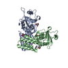
| Title | Staphylococcus aureus FusB bound to the small subunit of the S. aureus 70S ribosome (FusB-Sa70S:SSU) | |||||||||||||||
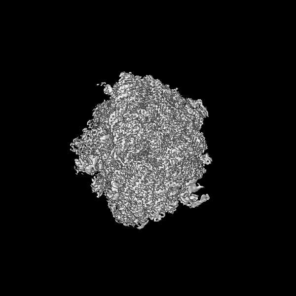
 Map data Map data | Local filtered map | |||||||||||||||
 Sample Sample |
| |||||||||||||||
 Keywords Keywords | RIBOSOME / fusidic acid / EF-G / antibiotic / FusB | |||||||||||||||
| Function / homology |  Function and homology information Function and homology informationlarge ribosomal subunit / transferase activity / ribosome biogenesis / ribosomal small subunit biogenesis / 5S rRNA binding / ribosomal large subunit assembly / small ribosomal subunit / small ribosomal subunit rRNA binding / cytosolic small ribosomal subunit / large ribosomal subunit rRNA binding ...large ribosomal subunit / transferase activity / ribosome biogenesis / ribosomal small subunit biogenesis / 5S rRNA binding / ribosomal large subunit assembly / small ribosomal subunit / small ribosomal subunit rRNA binding / cytosolic small ribosomal subunit / large ribosomal subunit rRNA binding / cytosolic large ribosomal subunit / cytoplasmic translation / tRNA binding / negative regulation of translation / rRNA binding / structural constituent of ribosome / ribosome / translation / ribonucleoprotein complex / mRNA binding / RNA binding / zinc ion binding / metal ion binding / cytoplasm / cytosol Similarity search - Function | |||||||||||||||
| Biological species |  Staphylococcus aureus subsp. aureus NCTC 8325 (bacteria) / Staphylococcus aureus subsp. aureus NCTC 8325 (bacteria) /  Staphylococcus aureus (bacteria) / Staphylococcus aureus (bacteria) / | |||||||||||||||
| Method | single particle reconstruction / cryo EM / Resolution: 2.22 Å | |||||||||||||||
 Authors Authors | Gonzalez-Lopez A / Selmer M | |||||||||||||||
| Funding support |  Sweden, 4 items Sweden, 4 items
| |||||||||||||||
 Citation Citation | Journal: Nat Commun / Year: 2025 Title: Structural mechanism of FusB-mediated rescue from fusidic acid inhibition of protein synthesis. Authors: Adrián González-López / Xueliang Ge / Daniel S D Larsson / Carina Sihlbom Wallem / Suparna Sanyal / Maria Selmer / Abstract: The antibiotic resistance protein FusB rescues protein synthesis from inhibition by fusidic acid (FA), which locks elongation factor G (EF-G) to the ribosome after GTP hydrolysis. Here, we present ...The antibiotic resistance protein FusB rescues protein synthesis from inhibition by fusidic acid (FA), which locks elongation factor G (EF-G) to the ribosome after GTP hydrolysis. Here, we present time-resolved single-particle cryo-EM structures explaining the mechanism of FusB-mediated rescue. FusB binds to the FA-trapped EF-G on the ribosome, causing large-scale conformational changes of EF-G that break interactions with the ribosome, tRNA, and mRNA. This leads to dissociation of EF-G from the ribosome, followed by FA release. We also observe two independent binding sites of FusB on the classical-state ribosome, overlapping with the binding site of EF-G to each of the ribosomal subunits, yet not inhibiting tRNA delivery. The affinity of FusB to the ribosome and the concentration of FusB in S. aureus during FusB-mediated resistance support that direct binding of FusB to ribosomes could occur in the cell. Our results reveal an intricate resistance mechanism involving specific interactions of FusB with both EF-G and the ribosome, and a non-canonical release pathway of EF-G. | |||||||||||||||
| History |
|
- Structure visualization
| Supplemental images |
|---|

- Downloads & links
-EMDB archive
| Map data | emd_51356.map.gz | 60.2 MB | EMDB map data format | |
|---|---|---|---|---|
| Header (meta data) | emd-51356-v30.xmlemd-51356.xml | 83.5 KB 83.5 KB | Display Display | EMDB header |
| FSC (resolution estimation) | emd_51356_fsc.xml | 22.3 KB | Display | FSC data file |
| Images |  emd_51356.png emd_51356.png | 104.4 KB | ||
| Masks | emd_51356_msk_1.map | 824 MB | Mask map | |
| Filedesc metadata | emd-51356.cif.gz | 16.1 KB | ||
| Others | emd_51356_additional_1.map.gzemd_51356_half_map_1.map.gzemd_51356_half_map_2.map.gz | 412.7 MB 763.4 MB 763.4 MB | ||
| Archive directory |  http://ftp.pdbj.org/pub/emdb/structures/EMD-51356ftp://ftp.pdbj.org/pub/emdb/structures/EMD-51356 http://ftp.pdbj.org/pub/emdb/structures/EMD-51356ftp://ftp.pdbj.org/pub/emdb/structures/EMD-51356 | HTTPS FTP |
-Related structure data
| Related structure data |  9ghgMC  9ghaC  9ghbC  9ghcC 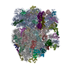 9ghdC  9gheC  9ghfC  9ghhC M: atomic model generated by this map C: citing same article ( |
|---|---|
| Similar structure data |


-Links
| EMDB pages | EMDB (EBI/PDBe) / EMDataResource |
|---|---|
| Related items in Molecule of the Month |


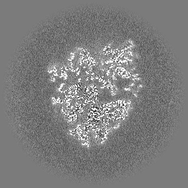
-Map
| File | Download / File: emd_51356.map.gz / Format: CCP4 / Size: 824 MB / Type: IMAGE STORED AS FLOATING POINT NUMBER (4 BYTES) | ||||||||||||||||||||||||||||||||||||
|---|---|---|---|---|---|---|---|---|---|---|---|---|---|---|---|---|---|---|---|---|---|---|---|---|---|---|---|---|---|---|---|---|---|---|---|---|---|
| Annotation | Local filtered map | ||||||||||||||||||||||||||||||||||||
| Projections & slices | Image control
Images are generated by Spider. | ||||||||||||||||||||||||||||||||||||
| Voxel size | X=Y=Z: 0.728 Å | ||||||||||||||||||||||||||||||||||||
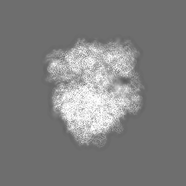
| Density |
| ||||||||||||||||||||||||||||||||||||
| Symmetry | Space group: 1 | ||||||||||||||||||||||||||||||||||||
| Details | EMDB XML:
|
 Z (Sec.)
 Y (Row.)
 X (Col.)


-Supplemental data
-Mask #1
| File | emd_51356_msk_1.map | ||||||||||||
|---|---|---|---|---|---|---|---|---|---|---|---|---|---|
| Projections & Slices |
| ||||||||||||
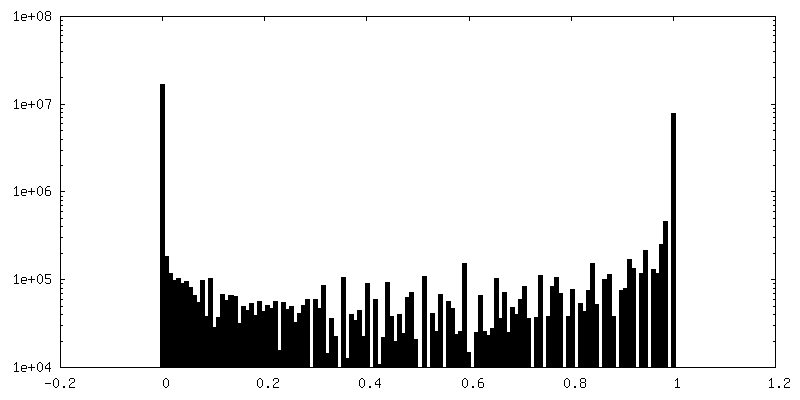
| Density Histograms |


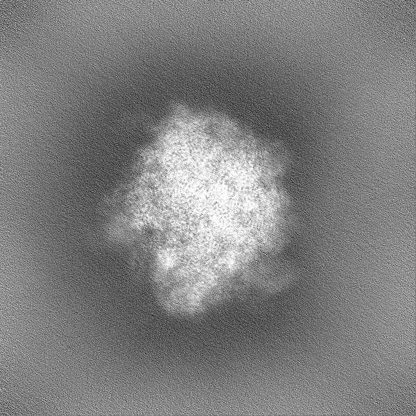
-Additional map: Unsharpened map
| File | emd_51356_additional_1.map | ||||||||||||
|---|---|---|---|---|---|---|---|---|---|---|---|---|---|
| Annotation | Unsharpened map | ||||||||||||
| Projections & Slices |
| ||||||||||||
| Density Histograms |


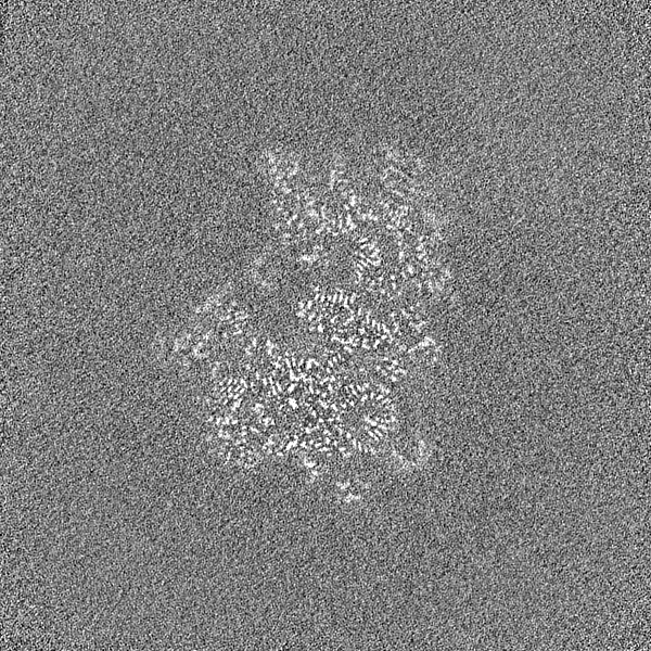
-Half map: Half map B
| File | emd_51356_half_map_1.map | ||||||||||||
|---|---|---|---|---|---|---|---|---|---|---|---|---|---|
| Annotation | Half map B | ||||||||||||
| Projections & Slices |
| ||||||||||||
| Density Histograms |


-Half map: Half map A
| File | emd_51356_half_map_2.map | ||||||||||||
|---|---|---|---|---|---|---|---|---|---|---|---|---|---|
| Annotation | Half map A | ||||||||||||
| Projections & Slices |
| ||||||||||||
| Density Histograms |


- Sample components
+Entire : FusB on 70S ribosomes
+Supramolecule #1: FusB on 70S ribosomes
+Supramolecule #2: FusB
+Macromolecule #1: 50S ribosomal protein L28
+Macromolecule #2: 50S ribosomal protein L29
+Macromolecule #3: 50S ribosomal protein L30
+Macromolecule #4: 50S ribosomal protein L31 type B
+Macromolecule #5: Large ribosomal subunit protein bL32
+Macromolecule #6: Large ribosomal subunit protein bL33A
+Macromolecule #7: 50S ribosomal protein L34
+Macromolecule #8: 50S ribosomal protein L35
+Macromolecule #9: 50S ribosomal protein L36
+Macromolecule #12: Far1
+Macromolecule #14: 50S ribosomal protein L2
+Macromolecule #15: 50S ribosomal protein L3
+Macromolecule #16: 50S ribosomal protein L4
+Macromolecule #17: 50S ribosomal protein L5
+Macromolecule #18: 50S ribosomal protein L6
+Macromolecule #19: 50S ribosomal protein L13
+Macromolecule #20: 50S ribosomal protein L14
+Macromolecule #21: 50S ribosomal protein L15
+Macromolecule #22: 50S ribosomal protein L16
+Macromolecule #23: 50S ribosomal protein L17
+Macromolecule #24: 50S ribosomal protein L18
+Macromolecule #25: 50S ribosomal protein L19
+Macromolecule #26: 50S ribosomal protein L20
+Macromolecule #27: 50S ribosomal protein L21
+Macromolecule #28: 50S ribosomal protein L22
+Macromolecule #29: 50S ribosomal protein L23
+Macromolecule #30: 50S ribosomal protein L24
+Macromolecule #31: 50S ribosomal protein L25
+Macromolecule #32: 50S ribosomal protein L27
+Macromolecule #35: 30S ribosomal protein S2
+Macromolecule #36: 30S ribosomal protein S3
+Macromolecule #37: 30S ribosomal protein S4
+Macromolecule #38: 30S ribosomal protein S5
+Macromolecule #39: 30S ribosomal protein S6
+Macromolecule #40: 30S ribosomal protein S7
+Macromolecule #41: 30S ribosomal protein S8
+Macromolecule #42: 30S ribosomal protein S9
+Macromolecule #43: Small ribosomal subunit protein uS10
+Macromolecule #44: 30S ribosomal protein S11
+Macromolecule #45: 30S ribosomal protein S12
+Macromolecule #46: 30S ribosomal protein S13
+Macromolecule #47: 30S ribosomal protein S14 type Z
+Macromolecule #48: 30S ribosomal protein S15
+Macromolecule #49: 30S ribosomal protein S16
+Macromolecule #50: 30S ribosomal protein S17
+Macromolecule #51: 30S ribosomal protein S18
+Macromolecule #52: 30S ribosomal protein S19
+Macromolecule #53: 30S ribosomal protein S20
+Macromolecule #54: 30S ribosomal protein S21
+Macromolecule #10: 23S rRNA
+Macromolecule #11: 5S rRNA
+Macromolecule #13: E-site tRNA
+Macromolecule #33: 23S rRNA
+Macromolecule #34: mRNA
+Macromolecule #55: ZINC ION
+Macromolecule #56: MAGNESIUM ION
+Macromolecule #57: 1,4-DIAMINOBUTANE
+Macromolecule #58: water

-Experimental details
-Structure determination
| Method | cryo EM |
|---|---|
 Processing Processing | single particle reconstruction |
| Aggregation state | particle |
-Sample preparation
| Buffer | pH: 7.5 Component:
| |||||||||||||||||||||||||||
|---|---|---|---|---|---|---|---|---|---|---|---|---|---|---|---|---|---|---|---|---|---|---|---|---|---|---|---|---|
| Grid | Model: Quantifoil R2/2 / Material: COPPER / Mesh: 300 / Support film - Material: CARBON / Support film - topology: CONTINUOUS / Support film - Film thickness: 2 / Pretreatment - Type: GLOW DISCHARGE / Pretreatment - Time: 30 sec. / Pretreatment - Atmosphere: AIR / Pretreatment - Pressure: 0.039 kPa | |||||||||||||||||||||||||||
| Vitrification | Cryogen name: ETHANE / Chamber humidity: 95 % / Chamber temperature: 277.15 K / Instrument: FEI VITROBOT MARK IV |
- Electron microscopy
| Microscope | TFS KRIOS |
|---|---|
| Specialist optics | Energy filter - Name: TFS Selectris / Energy filter - Slit width: 10 eV |
| Image recording | Film or detector model: TFS FALCON 4i (4k x 4k) / Number grids imaged: 1 / Number real images: 10064 / Average exposure time: 2.14 sec. / Average electron dose: 28.14 e/Å2 |
| Electron beam | Acceleration voltage: 300 kV / Electron source:  FIELD EMISSION GUN FIELD EMISSION GUN |
| Electron optics | C2 aperture diameter: 50.0 µm / Illumination mode: FLOOD BEAM / Imaging mode: BRIGHT FIELD / Cs: 2.7 mm / Nominal defocus max: 1.3 µm / Nominal defocus min: 0.7000000000000001 µm / Nominal magnification: 165000 |
| Sample stage | Specimen holder model: FEI TITAN KRIOS AUTOGRID HOLDER / Cooling holder cryogen: NITROGEN |
| Experimental equipment |  Model: Titan Krios / Image courtesy: FEI Company |
+Image processing

-Atomic model buiding 1
| Initial model |
| ||||||||||||
|---|---|---|---|---|---|---|---|---|---|---|---|---|---|
| Refinement | Space: REAL | ||||||||||||
| Output model | PDB-9ghg: |