Movie
Movie Controller

[English] 日本語
 Yorodumi
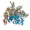
- EMDB-51258: RNAP-TopoI complex on duplex scaffold - focused map (RNAP beta pr... -
+ Open data

- Basic information
| Entry |  | |||||||||
|---|---|---|---|---|---|---|---|---|---|---|
| Title | RNAP-TopoI complex on duplex scaffold - focused map (RNAP beta protrusion and lobe) | |||||||||
 Map data Map data | RNAP-TopoI complex on duplex scaffold focused refinement (RNAP beta protrusion & lobe) - deepemhanced map | |||||||||
 Sample Sample |
| |||||||||
 Keywords Keywords | RNA polymerase / topoisomerase / DNA topology / TRANSCRIPTION | |||||||||
| Biological species |  | |||||||||
| Method | single particle reconstruction / cryo EM / Resolution: 2.8 Å | |||||||||
 Authors Authors | Vidmar V / Weixlbaumer A / Lamour V | |||||||||
| Funding support |  France, 2 items France, 2 items
| |||||||||
 Citation Citation | Journal: Nat Struct Mol Biol / Year: 2026 Title: DNA topoisomerase I acts as supercoiling sensor for bacterial transcription elongation. Authors: Vita Vidmar / Céline Borde / Lisa Bruno / Nataliya Miropolskaya / Maria Takacs / Claire Batisse / Charlotte Saint-André / Chengjin Zhu / Olivier Espéli / Valérie Lamour / Albert Weixlbaumer / Abstract: During transcription, RNA polymerase (RNAP) continuously unwinds and rewinds DNA, generating negative and positive supercoils upstream and downstream, respectively. Using single-particle cryo-EM, we ...During transcription, RNA polymerase (RNAP) continuously unwinds and rewinds DNA, generating negative and positive supercoils upstream and downstream, respectively. Using single-particle cryo-EM, we elucidated how bacterial RNAP and DNA topoisomerase I (TopoI), which relaxes negative supercoils, operate in close spatial proximity. TopoI binds to relaxed DNA upstream of RNAP, and this involves a conformational switch in the TopoI functional domains. This suggests that TopoI exerts a sensing role before the formation of negative supercoils. On DNA substrates mimicking negatively supercoiled DNA, TopoI threads one strand into the active site for cleavage and binds the complementary strand with an auxiliary domain. Transcriptomic and phenotypic analyses suggest that mutations affecting conformational changes in TopoI impact gene expression and operon polarity in bacteria. In summary, we propose a comprehensive model for DNA relaxation in the proximity of active bacterial transcription. | |||||||||
| History |
|
- Structure visualization
| Supplemental images |
|---|

- Downloads & links
-EMDB archive
| Map data | emd_51258.map.gz | 146.1 MB |  EMDB map data format EMDB map data format | |
|---|---|---|---|---|
| Header (meta data) | emd-51258-v30.xmlemd-51258.xml | 24.4 KB 24.4 KB | Display Display | EMDB header |
| FSC (resolution estimation) | emd_51258_fsc.xml | 11.9 KB | Display | FSC data file |
| Images |  emd_51258.png emd_51258.png | 41.5 KB | ||
| Masks | emd_51258_msk_1.mapemd_51258_msk_2.map | 178 MB 178 MB | Mask map | |
| Filedesc metadata | emd-51258.cif.gz | 5.2 KB | ||
| Others | emd_51258_additional_1.map.gzemd_51258_half_map_1.map.gzemd_51258_half_map_2.map.gz | 86 MB 163.7 MB 163.7 MB | ||
| Archive directory |  http://ftp.pdbj.org/pub/emdb/structures/EMD-51258ftp://ftp.pdbj.org/pub/emdb/structures/EMD-51258 http://ftp.pdbj.org/pub/emdb/structures/EMD-51258ftp://ftp.pdbj.org/pub/emdb/structures/EMD-51258 | HTTPS FTP |
-Related structure data


-Links
| EMDB pages | EMDB (EBI/PDBe) / EMDataResource |
|---|
-Map
| File | Download / File: emd_51258.map.gz / Format: CCP4 / Size: 178 MB / Type: IMAGE STORED AS FLOATING POINT NUMBER (4 BYTES) | ||||||||||||||||||||||||||||||||||||
|---|---|---|---|---|---|---|---|---|---|---|---|---|---|---|---|---|---|---|---|---|---|---|---|---|---|---|---|---|---|---|---|---|---|---|---|---|---|
| Annotation | RNAP-TopoI complex on duplex scaffold focused refinement (RNAP beta protrusion & lobe) - deepemhanced map | ||||||||||||||||||||||||||||||||||||
| Projections & slices | Image control
Images are generated by Spider. | ||||||||||||||||||||||||||||||||||||
| Voxel size | X=Y=Z: 0.839 Å | ||||||||||||||||||||||||||||||||||||
| Density |
| ||||||||||||||||||||||||||||||||||||
| Symmetry | Space group: 1 | ||||||||||||||||||||||||||||||||||||
| Details | EMDB XML:
|
 Z (Sec.)
 Y (Row.)
 X (Col.)


-Supplemental data
-Mask #1
| File | emd_51258_msk_1.map | ||||||||||||
|---|---|---|---|---|---|---|---|---|---|---|---|---|---|
| Projections & Slices |
| ||||||||||||
| Density Histograms |


-Mask #2
| File | emd_51258_msk_2.map | ||||||||||||
|---|---|---|---|---|---|---|---|---|---|---|---|---|---|
| Projections & Slices |
| ||||||||||||
| Density Histograms |


-Additional map: RNAP-TopoI complex on duplex scaffold focused refinement (RNAP...
| File | emd_51258_additional_1.map | ||||||||||||
|---|---|---|---|---|---|---|---|---|---|---|---|---|---|
| Annotation | RNAP-TopoI complex on duplex scaffold focused refinement (RNAP beta protrusion & lobe) - unsharpened map | ||||||||||||
| Projections & Slices |
| ||||||||||||
| Density Histograms |


-Half map: RNAP-TopoI complex on duplex scaffold focused refinement (RNAP...
| File | emd_51258_half_map_1.map | ||||||||||||
|---|---|---|---|---|---|---|---|---|---|---|---|---|---|
| Annotation | RNAP-TopoI complex on duplex scaffold focused refinement (RNAP beta protrusion & lobe) - halfmap A | ||||||||||||
| Projections & Slices |
| ||||||||||||
| Density Histograms |


-Half map: RNAP-TopoI complex on duplex scaffold focused refinement (RNAP...
| File | emd_51258_half_map_2.map | ||||||||||||
|---|---|---|---|---|---|---|---|---|---|---|---|---|---|
| Annotation | RNAP-TopoI complex on duplex scaffold focused refinement (RNAP beta protrusion & lobe) - halfmap B | ||||||||||||
| Projections & Slices |
| ||||||||||||
| Density Histograms |


- Sample components
-Entire : RNAP-TopoI complex on duplex scaffold
| Entire | Name: RNAP-TopoI complex on duplex scaffold |
|---|---|
| Components |
|
-Supramolecule #1: RNAP-TopoI complex on duplex scaffold
| Supramolecule | Name: RNAP-TopoI complex on duplex scaffold / type: complex / ID: 1 / Parent: 0 / Macromolecule list: #1-#9 |
|---|---|
| Molecular weight | Theoretical: 100 KDa |
-Supramolecule #2: DNA-directed RNA polymerase elongation complex
| Supramolecule | Name: DNA-directed RNA polymerase elongation complex / type: complex / ID: 2 / Parent: 1 / Macromolecule list: #1-#5, #7-#9 |
|---|---|
| Source (natural) | Organism: |
-Supramolecule #3: DNA topoisomerase 1
| Supramolecule | Name: DNA topoisomerase 1 / type: complex / ID: 3 / Parent: 1 / Macromolecule list: #6 |
|---|---|
| Source (natural) | Organism: |
-Experimental details
-Structure determination
| Method | cryo EM |
|---|---|
 Processing Processing | single particle reconstruction |
| Aggregation state | particle |
-Sample preparation
| Concentration | 10 mg/mL | ||||||||||||
|---|---|---|---|---|---|---|---|---|---|---|---|---|---|
| Buffer | pH: 7.5 Component:
| ||||||||||||
| Grid | Model: UltrAuFoil R1.2/1.3 / Material: GOLD / Mesh: 300 / Support film - Material: GOLD / Support film - topology: HOLEY / Support film - Film thickness: 50 / Pretreatment - Type: PLASMA CLEANING / Pretreatment - Time: 35 sec. / Pretreatment - Atmosphere: OTHER | ||||||||||||
| Vitrification | Cryogen name: ETHANE / Chamber humidity: 100 % / Chamber temperature: 283 K / Instrument: FEI VITROBOT MARK IV |
- Electron microscopy
| Microscope | TFS KRIOS |
|---|---|
| Image recording | Film or detector model: GATAN K3 (6k x 4k) / Average electron dose: 56.6 e/Å2 |
| Electron beam | Acceleration voltage: 300 kV / Electron source:  FIELD EMISSION GUN FIELD EMISSION GUN |
| Electron optics | Illumination mode: FLOOD BEAM / Imaging mode: BRIGHT FIELD / Nominal defocus max: 2.5 µm / Nominal defocus min: 0.8 µm |
| Sample stage | Cooling holder cryogen: NITROGEN |
| Experimental equipment |  Model: Titan Krios / Image courtesy: FEI Company |
+Image processing

-Atomic model buiding 1
| Initial model |
| ||||||
|---|---|---|---|---|---|---|---|
| Details | Initial rigid body fit was done in UCSF Chimera, followed by flexible fitting in Isolde. The entire model was further refined in Phenix and Coot. | ||||||
| Refinement | Space: REAL |