 Movie
Movie Controller


+ Open data

- Basic information
| Entry |  | |||||||||
|---|---|---|---|---|---|---|---|---|---|---|
| Title | Cryo-EM structure of CdaA-DAC domain in complex with GlmM | |||||||||
 Map data Map data | ||||||||||
 Sample Sample |
| |||||||||
 Keywords Keywords | cyclic-di-AMP cyclace / Inhibitor / Complex / PROTEIN BINDING | |||||||||
| Function / homology |  Function and homology information Function and homology informationphosphoglucosamine mutase / phosphoglucosamine mutase activity / phosphomannomutase activity / diadenylate cyclase / diadenylate cyclase activity / UDP-N-acetylglucosamine biosynthetic process / cAMP biosynthetic process / adenylate cyclase activity / peptidoglycan biosynthetic process / carbohydrate metabolic process ...phosphoglucosamine mutase / phosphoglucosamine mutase activity / phosphomannomutase activity / diadenylate cyclase / diadenylate cyclase activity / UDP-N-acetylglucosamine biosynthetic process / cAMP biosynthetic process / adenylate cyclase activity / peptidoglycan biosynthetic process / carbohydrate metabolic process / magnesium ion binding / ATP binding / plasma membrane / cytosol Similarity search - Function | |||||||||
| Biological species |  Lactococcus lactis subsp. lactis (lactic acid bacteria) Lactococcus lactis subsp. lactis (lactic acid bacteria) | |||||||||
| Method | single particle reconstruction / cryo EM / Resolution: 4.84 Å | |||||||||
 Authors Authors | Drougkas P / Paulino C / Poolman B | |||||||||
| Funding support |  Netherlands, 1 items Netherlands, 1 items
| |||||||||
 Citation Citation | Journal: Commun Biol / Year: 2024 Title: Membrane-embedded CdaA is required for efficient synthesis of second messenger cyclic di-AMP. Authors: Alexander J Foster / Haoyang Li / Panagiotis Drougkas / Gea K Schuurman-Wolters / Joeri Ten Kate / Cristina Paulino / Bert Poolman /  Abstract: Cyclic di-adenylate monophosphate (cyclic di-AMP) is an important second messenger in microorganisms. Cyclic di-AMP regulates bacterial cell volume and turgor via control of potassium and compatible ...Cyclic di-adenylate monophosphate (cyclic di-AMP) is an important second messenger in microorganisms. Cyclic di-AMP regulates bacterial cell volume and turgor via control of potassium and compatible solute transport but is also involved in many other processes, including the activation of the metazoan innate immune response to bacterial infections. We compare the activity of full-length membrane-embedded CdaA, the enzyme that synthesizes cyclic di-AMP, with the water-soluble catalytic domain CdaA-DAC. Purified CdaA from L. lactis was studied in the detergent-solubilized state, and in lipid nanodiscs and vesicles. We show that CdaA is tetrameric and the membrane-bound complex has more than 2-orders of magnitude higher activity than soluble CdaA-DAC. CdaA activity increases with pH but does not strongly depend on the salt or lipid content, factors that are crucial for the control of osmoregulatory transporters. Cryo-EM and in-silico structure prediction of CdaA show that the two DAC dimers engage in a head-to-head interaction, leading to cyclic-di-AMP formation. The inhibitor phosphoglucomutase prevents this active conformation. We observe dynamic flexibility between the catalytic and membrane domains, even in the presence of ATP or non-hydrolyzable substrate ApCpp. This is the first comprehensive functional and structural characterization of a full-length cyclic di-AMP-specific cyclase. | |||||||||
| History |
|
- Structure visualization
| Supplemental images |
|---|

- Downloads & links
-EMDB archive
| Map data | emd_51096.map.gz | 59.7 MB | EMDB map data format | |
|---|---|---|---|---|
| Header (meta data) | emd-51096-v30.xmlemd-51096.xml | 20.4 KB 20.4 KB | Display Display | EMDB header |
| FSC (resolution estimation) | emd_51096_fsc.xml | 9.2 KB | Display | FSC data file |
| Images |  emd_51096.png emd_51096.png | 109.5 KB | ||
| Masks | emd_51096_msk_1.map | 64 MB | Mask map | |
| Filedesc metadata | emd-51096.cif.gz | 6.7 KB | ||
| Others | emd_51096_half_map_1.map.gzemd_51096_half_map_2.map.gz | 49.7 MB 49.7 MB | ||
| Archive directory |  http://ftp.pdbj.org/pub/emdb/structures/EMD-51096ftp://ftp.pdbj.org/pub/emdb/structures/EMD-51096 http://ftp.pdbj.org/pub/emdb/structures/EMD-51096ftp://ftp.pdbj.org/pub/emdb/structures/EMD-51096 | HTTPS FTP |
-Validation report
| Summary document | emd_51096_validation.pdf.gz | 846.7 KB | Display | EMDB validaton report |
|---|---|---|---|---|
| Full document | emd_51096_full_validation.pdf.gz | 846.3 KB | Display | |
| Data in XML | emd_51096_validation.xml.gz | 15.9 KB | Display | |
| Data in CIF | emd_51096_validation.cif.gz | 21.1 KB | Display | |
| Arichive directory | https://ftp.pdbj.org/pub/emdb/validation_reports/EMD-51096ftp://ftp.pdbj.org/pub/emdb/validation_reports/EMD-51096 | HTTPS FTP |
-Related structure data
| Related structure data | 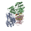 9g69MC M: atomic model generated by this map C: citing same article ( |
|---|---|
| Similar structure data |
-Links
| EMDB pages | EMDB (EBI/PDBe) / EMDataResource |
|---|
-Map
| File | Download / File: emd_51096.map.gz / Format: CCP4 / Size: 64 MB / Type: IMAGE STORED AS FLOATING POINT NUMBER (4 BYTES) | ||||||||||||||||||||||||||||||||||||
|---|---|---|---|---|---|---|---|---|---|---|---|---|---|---|---|---|---|---|---|---|---|---|---|---|---|---|---|---|---|---|---|---|---|---|---|---|---|
| Projections & slices | Image control
Images are generated by Spider. | ||||||||||||||||||||||||||||||||||||
| Voxel size | X=Y=Z: 1.022 Å | ||||||||||||||||||||||||||||||||||||
| Density |
| ||||||||||||||||||||||||||||||||||||
| Symmetry | Space group: 1 | ||||||||||||||||||||||||||||||||||||
| Details | EMDB XML:
|
 Z (Sec.)
 Y (Row.)
 X (Col.)


-Supplemental data
-Mask #1
| File | emd_51096_msk_1.map | ||||||||||||
|---|---|---|---|---|---|---|---|---|---|---|---|---|---|
| Projections & Slices |
| ||||||||||||
| Density Histograms |


-Half map: #2
| File | emd_51096_half_map_1.map | ||||||||||||
|---|---|---|---|---|---|---|---|---|---|---|---|---|---|
| Projections & Slices |
| ||||||||||||
| Density Histograms |


-Half map: #1
| File | emd_51096_half_map_2.map | ||||||||||||
|---|---|---|---|---|---|---|---|---|---|---|---|---|---|
| Projections & Slices |
| ||||||||||||
| Density Histograms |


- Sample components
-Entire : Diadenylate cyclase CdaA, Phosphoglucosamine mutase GlmM, ATP
| Entire | Name: Diadenylate cyclase CdaA, Phosphoglucosamine mutase GlmM, ATP |
|---|---|
| Components |
|
-Supramolecule #1: Diadenylate cyclase CdaA, Phosphoglucosamine mutase GlmM, ATP
| Supramolecule | Name: Diadenylate cyclase CdaA, Phosphoglucosamine mutase GlmM, ATP type: complex / ID: 1 / Parent: 0 / Macromolecule list: #1-#2 |
|---|---|
| Source (natural) | Organism: Lactococcus lactis subsp. lactis (lactic acid bacteria) |
-Macromolecule #1: Phosphoglucosamine mutase
| Macromolecule | Name: Phosphoglucosamine mutase / type: protein_or_peptide / ID: 1 / Number of copies: 2 / Enantiomer: LEVO / EC number: phosphoglucosamine mutase |
|---|---|
| Source (natural) | Organism: Lactococcus lactis subsp. lactis (lactic acid bacteria) |
| Molecular weight | Theoretical: 48.521879 KDa |
| Recombinant expression | Organism: Lactococcus lactis subsp. lactis (lactic acid bacteria) |
| Sequence | String: MGKYFGTDGV RGEANVELTP EMAFKLGRFG GYVLSQHELG TPKVYVGRDT RISGQMLASS LISGLLSVGI EVYDLGVIAT PGVAYLVKK DGASAGVMIS ASHNPALDNG IKFFGGDGYK LEDEKELEIE ALIDAEEDTL PRPSAQGLGM LHDYIEGVRK Y QAFLKTTA ...String: MGKYFGTDGV RGEANVELTP EMAFKLGRFG GYVLSQHELG TPKVYVGRDT RISGQMLASS LISGLLSVGI EVYDLGVIAT PGVAYLVKK DGASAGVMIS ASHNPALDNG IKFFGGDGYK LEDEKELEIE ALIDAEEDTL PRPSAQGLGM LHDYIEGVRK Y QAFLKTTA EGNFEGYKVV LDTANGAAYT SARAVFADLE ANLTVIGENP DGLNINVKVG STHPEAMAKK VVETGSDLGL AF DGDADRL IAVDENGEIV DGDKIMFIVG KYLLGQGKLA QDTLVTTVMS NLGFHLALEE AGINSVITAV GDRYVVEEMK KNN YNFGGE QSGHMIFLDY NTTGDGQLSA IQLLKVMRET GKSLSELASE VTIYPQKLVN VRVKDNAAKK SAMDVPAIQK VISE METSM NGKGRILVRP SGTEPLLRVM AEAPTHEEVN HVVDTIVEVV EAEIGVK UniProtKB: Phosphoglucosamine mutase |
-Macromolecule #2: Diadenylate cyclase
| Macromolecule | Name: Diadenylate cyclase / type: protein_or_peptide / ID: 2 / Number of copies: 2 / Enantiomer: LEVO / EC number: diadenylate cyclase |
|---|---|
| Source (natural) | Organism: Lactococcus lactis subsp. lactis (lactic acid bacteria) |
| Molecular weight | Theoretical: 32.433725 KDa |
| Recombinant expression | Organism: Lactococcus lactis subsp. lactis (lactic acid bacteria) |
| Sequence | String: MTDFNQFFNL EFWQKIFELN ESPLRIAIAV LDIAIVSFFL YQAIRFVQGT KLMTLVRGVI IFIFIRIIAG LIGLTTVEWL LNQVITYGV IAGVIIFQPE IRRALESLGR TTTLFTPTKK GSLDGHISAY EKSFAYMSER KIGALIAIER GQNLNEFVST G IRLDADIT ...String: MTDFNQFFNL EFWQKIFELN ESPLRIAIAV LDIAIVSFFL YQAIRFVQGT KLMTLVRGVI IFIFIRIIAG LIGLTTVEWL LNQVITYGV IAGVIIFQPE IRRALESLGR TTTLFTPTKK GSLDGHISAY EKSFAYMSER KIGALIAIER GQNLNEFVST G IRLDADIT SELLINIFIP NTPLHDGAVI VQGNKIAVTS AYLPLTEKSG ISKQFGTRHR AAIGLSEVSD ALILVVSEET GG ISVAHNG EFFADISKEK FHDILVAILV PKVEKTVRGT GRIRKNKVEE KKNGK UniProtKB: Diadenylate cyclase |
-Macromolecule #3: ADENOSINE-5'-TRIPHOSPHATE
| Macromolecule | Name: ADENOSINE-5'-TRIPHOSPHATE / type: ligand / ID: 3 / Number of copies: 1 / Formula: ATP |
|---|---|
| Molecular weight | Theoretical: 507.181 Da |
| Chemical component information |  ChemComp-ATP: |
-Experimental details
-Structure determination
| Method | cryo EM |
|---|---|
 Processing Processing | single particle reconstruction |
| Aggregation state | particle |
-Sample preparation
| Concentration | 1 mg/mL | |||||||||
|---|---|---|---|---|---|---|---|---|---|---|
| Buffer | pH: 7 Component:
| |||||||||
| Grid | Model: Quantifoil R1.2/1.3 / Material: GOLD / Mesh: 300 / Support film - Material: CARBON / Support film - topology: HOLEY / Pretreatment - Type: GLOW DISCHARGE / Pretreatment - Time: 20 sec. / Details: 5 mA | |||||||||
| Vitrification | Cryogen name: ETHANE-PROPANE / Chamber humidity: 100 % / Chamber temperature: 293 K / Instrument: FEI VITROBOT MARK IV Details: 13.2 uM of GlmM and 10 mM of Mn-ATP were added prior to sample application onto the grids. Grids were blotted for 4 seconds.. | |||||||||
| Details | Full-length CdaA |
- Electron microscopy
| Microscope | FEI TALOS ARCTICA |
|---|---|
| Temperature | Min: 90.0 K / Max: 105.0 K |
| Specialist optics | Energy filter - Name: GIF Bioquantum / Energy filter - Slit width: 20 eV |
| Image recording | Film or detector model: GATAN K2 SUMMIT (4k x 4k) / Detector mode: COUNTING / Digitization - Dimensions - Width: 3838 pixel / Digitization - Dimensions - Height: 3710 pixel / Digitization - Frames/image: 1-60 / Number grids imaged: 1 / Number real images: 5150 / Average exposure time: 9.0 sec. / Average electron dose: 53.6 e/Å2 |
| Electron beam | Acceleration voltage: 200 kV / Electron source:  FIELD EMISSION GUN FIELD EMISSION GUN |
| Electron optics | C2 aperture diameter: 100.0 µm / Calibrated defocus max: 2.0 µm / Calibrated defocus min: 0.5 µm / Calibrated magnification: 49407 / Illumination mode: FLOOD BEAM / Imaging mode: BRIGHT FIELD / Cs: 2.7 mm / Nominal defocus max: 2.0 µm / Nominal defocus min: 0.5 µm / Nominal magnification: 130000 |
| Sample stage | Specimen holder model: FEI TITAN KRIOS AUTOGRID HOLDER / Cooling holder cryogen: NITROGEN |
| Experimental equipment |  Model: Talos Arctica / Image courtesy: FEI Company |
+Image processing

-Atomic model buiding 1
| Initial model | Chain - Source name: AlphaFold / Chain - Initial model type: in silico model / Details: v3 |
|---|---|
| Refinement | Space: REAL |
| Output model | PDB-9g69: |
