Journal: Acta Crystallogr F Struct Biol Commun / Year: 2024 Title: Revisiting sodium phosphotungstate and ammonium molybdate as nonradioactive negative-staining agents for single-particle analysis. Authors: Monika Gunkel / Arthur Macha / Elmar Behrmann / Abstract: This study reports the successful replacement of uranyl-based stains by either sodium phosphotungstate or ammonium molybdate in negative-staining electron microscopy. Using apoferritin as a test ...This study reports the successful replacement of uranyl-based stains by either sodium phosphotungstate or ammonium molybdate in negative-staining electron microscopy. Using apoferritin as a test specimen, it is demonstrated that in combination with a facile on-grid fixation step, both stains yield comparable images to uranyl formate. Subsequently, using β-galactosidase, it is shown that both stains can also successfully be employed for single-particle analysis, yielding virtually indistinguishable results from uranyl formate. As both replacement stains are nonradioactive, they are not subjected to the same handling restrictions as uranyl-based stains. Therefore they are not only cheaper to use, but also make decentralized sample-grid preparation, directly after purification, accessible to a broader range of scientists.
Details: 25 mM Tris at pH 8, 50 mM NaCl, 2 mM MgCl2, and 0.5 mM TCEP
Staining
Type: NEGATIVE / Material: Uranyl Formate Details: Negative stained EM specimens were prepared using uranyl formate negative staining.
Grid
Model: Quantifoil / Material: COPPER / Mesh: 200 / Support film - Material: CARBON / Support film - topology: CONTINUOUS / Pretreatment - Type: GLOW DISCHARGE / Pretreatment - Time: 30 sec. / Pretreatment - Atmosphere: AIR
Details
This sample was monodisperse
-
Electron microscopy
Microscope
TFS TALOS L120C
Details
Preliminary grid screening was performed manually.
Image recording
Film or detector model: FEI CETA (4k x 4k) / Number grids imaged: 1 / Number real images: 200 / Average exposure time: 1.0 sec. / Average electron dose: 33.0 e/Å2 / Details: 163 were used
Electron beam
Acceleration voltage: 120 kV / Electron source: LAB6
In the structure databanks used in Yorodumi, some data are registered as the other names, "COVID-19 virus" and "2019-nCoV". Here are the details of the virus and the list of structure data.
Jan 31, 2019. EMDB accession codes are about to change! (news from PDBe EMDB page)
EMDB accession codes are about to change! (news from PDBe EMDB page)
The allocation of 4 digits for EMDB accession codes will soon come to an end. Whilst these codes will remain in use, new EMDB accession codes will include an additional digit and will expand incrementally as the available range of codes is exhausted. The current 4-digit format prefixed with “EMD-” (i.e. EMD-XXXX) will advance to a 5-digit format (i.e. EMD-XXXXX), and so on. It is currently estimated that the 4-digit codes will be depleted around Spring 2019, at which point the 5-digit format will come into force.
The EM Navigator/Yorodumi systems omit the EMD- prefix.
Related info.:Q: What is EMD? / ID/Accession-code notation in Yorodumi/EM Navigator
Yorodumi is a browser for structure data from EMDB, PDB, SASBDB, etc.
This page is also the successor to EM Navigator detail page, and also detail information page/front-end page for Omokage search.
The word "yorodu" (or yorozu) is an old Japanese word meaning "ten thousand". "mi" (miru) is to see.
Related info.:EMDB / PDB / SASBDB / Comparison of 3 databanks / Yorodumi Search / Aug 31, 2016. New EM Navigator & Yorodumi / Yorodumi Papers / Jmol/JSmol / Function and homology information / Changes in new EM Navigator and Yorodumi
 Movie
Movie Controller


 Open data
 Basic information
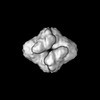
 Map data
 Sample
 Keywords
 Function and homology information
 Authors
 Germany, 1 items
  Citation
 Structure visualization
 Downloads & links
 emd_51069.png
 http://ftp.pdbj.org/pub/emdb/structures/EMD-51069


 Z (Sec.)
 Y (Row.)
 X (Col.)


 Sample components
 Processing
 Electron microscopy