 Movie
Movie Controller

[English] 日本語
 Yorodumi
- EMDB-50702: E. coli 70S ribosome in situ structure for lamellae backside dama... -
+ Open data

- Basic information
| Entry |  | ||||||||||||
|---|---|---|---|---|---|---|---|---|---|---|---|---|---|
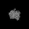
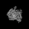
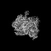
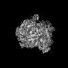
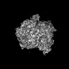
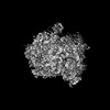
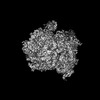
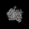
| Title | E. coli 70S ribosome in situ structure for lamellae backside damage analysis: 3 - 4 microns from lamellae backside | ||||||||||||
 Map data Map data | |||||||||||||
 Sample Sample |
| ||||||||||||
 Keywords Keywords | 70S E. coli ribosome / RIBOSOME | ||||||||||||
| Biological species |  | ||||||||||||
| Method | subtomogram averaging / cryo EM / Resolution: 6.6 Å | ||||||||||||
 Authors Authors | Watson H / Berger C / Grange M | ||||||||||||
| Funding support |  United Kingdom, 3 items United Kingdom, 3 items
| ||||||||||||
 Citation Citation | Journal: Nat Commun / Year: 2025 Title: Xenon plasma focused ion beam lamella fabrication on high-pressure frozen specimens for structural cell biology. Authors: Casper Berger / Helena Watson / James H Naismith / Maud Dumoux / Michael Grange / Abstract: Cryo focused ion beam lamella preparation is a potent tool for in situ structural biology, enabling the study of macromolecules in their native cellular environments. However, throughput is currently ...Cryo focused ion beam lamella preparation is a potent tool for in situ structural biology, enabling the study of macromolecules in their native cellular environments. However, throughput is currently limited, especially for thicker, more biologically complex samples. We describe how xenon plasma focused ion beam milling can be used for routine bulk milling of thicker, high-pressure frozen samples. We demonstrate lamellae preparation with a high success rate on these samples and determine a 4.0 Å structure of the Escherichia coli ribosome on these lamellae using sub volume averaging. We determine the effects on sample integrity of increased ion currents up to 60 nA during bulk milling of thicker planar samples, showing no measurable damage to macromolecules beyond an amorphous layer on the backside of the lamellae. The use of xenon results in substantial structural damage to particles up to approximately 30 nm in depth from the milled surfaces, and the effects of damage become negligibly small by 45 nm. Our results outline how the use of high currents using xenon plasma focused ion beam milling may be integrated into FIB milling regimes for preparing thin lamellae for high-resolution in situ structural biology. | ||||||||||||
| History |
|
- Structure visualization
| Supplemental images |
|---|

- Downloads & links
-EMDB archive
| Map data | emd_50702.map.gz | 33.1 MB |  EMDB map data format EMDB map data format | |
|---|---|---|---|---|
| Header (meta data) | emd-50702-v30.xmlemd-50702.xml | 14.2 KB 14.2 KB | Display Display | EMDB header |
| FSC (resolution estimation) | emd_50702_fsc.xml | 8 KB | Display | FSC data file |
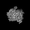
| Images |  emd_50702.png emd_50702.png | 101.6 KB | ||
| Masks | emd_50702_msk_1.map | 42.9 MB | Mask map | |
| Filedesc metadata | emd-50702.cif.gz | 4.3 KB | ||
| Others | emd_50702_half_map_1.map.gzemd_50702_half_map_2.map.gz | 33.2 MB 33.1 MB | ||
| Archive directory |  http://ftp.pdbj.org/pub/emdb/structures/EMD-50702ftp://ftp.pdbj.org/pub/emdb/structures/EMD-50702 http://ftp.pdbj.org/pub/emdb/structures/EMD-50702ftp://ftp.pdbj.org/pub/emdb/structures/EMD-50702 | HTTPS FTP |
-Validation report
| Summary document | emd_50702_validation.pdf.gz | 1.2 MB | Display | EMDB validaton report |
|---|---|---|---|---|
| Full document | emd_50702_full_validation.pdf.gz | 1.2 MB | Display | |
| Data in XML | emd_50702_validation.xml.gz | 14.1 KB | Display | |
| Data in CIF | emd_50702_validation.cif.gz | 19.6 KB | Display | |
| Arichive directory | https://ftp.pdbj.org/pub/emdb/validation_reports/EMD-50702ftp://ftp.pdbj.org/pub/emdb/validation_reports/EMD-50702 | HTTPS FTP |
-Related structure data
| Related structure data | C: citing same article ( |
|---|


-Links
| EMDB pages | EMDB (EBI/PDBe) / EMDataResource |
|---|
-Map
| File | Download / File: emd_50702.map.gz / Format: CCP4 / Size: 42.9 MB / Type: IMAGE STORED AS FLOATING POINT NUMBER (4 BYTES) | ||||||||||||||||||||||||||||||||||||
|---|---|---|---|---|---|---|---|---|---|---|---|---|---|---|---|---|---|---|---|---|---|---|---|---|---|---|---|---|---|---|---|---|---|---|---|---|---|
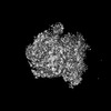
| Projections & slices | Image control
Images are generated by Spider. | ||||||||||||||||||||||||||||||||||||
| Voxel size | X=Y=Z: 1.9 Å | ||||||||||||||||||||||||||||||||||||
| Density |
| ||||||||||||||||||||||||||||||||||||
| Symmetry | Space group: 1 | ||||||||||||||||||||||||||||||||||||
| Details | EMDB XML:
|
 Z (Sec.)
 Y (Row.)
 X (Col.)


-Supplemental data
-Mask #1
| File | emd_50702_msk_1.map | ||||||||||||
|---|---|---|---|---|---|---|---|---|---|---|---|---|---|
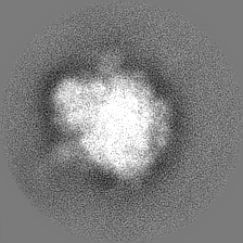
| Projections & Slices |
| ||||||||||||
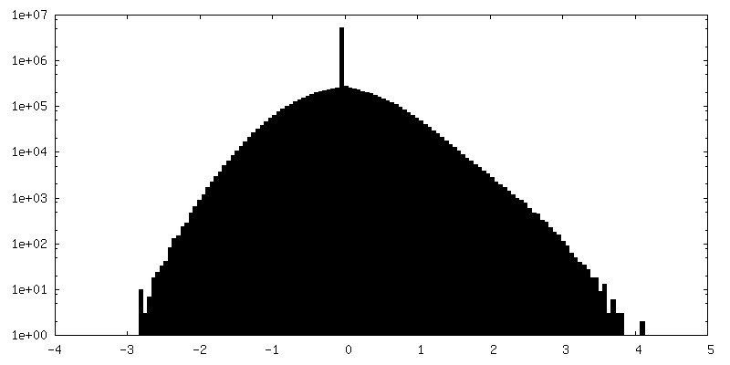
| Density Histograms |


-Half map: #1
| File | emd_50702_half_map_1.map | ||||||||||||
|---|---|---|---|---|---|---|---|---|---|---|---|---|---|
| Projections & Slices |
| ||||||||||||
| Density Histograms |


-Half map: #2
| File | emd_50702_half_map_2.map | ||||||||||||
|---|---|---|---|---|---|---|---|---|---|---|---|---|---|
| Projections & Slices |
| ||||||||||||
| Density Histograms |


- Sample components
-Entire : E. coli 70S ribosome
| Entire | Name: E. coli 70S ribosome |
|---|---|
| Components |
|
-Supramolecule #1: E. coli 70S ribosome
| Supramolecule | Name: E. coli 70S ribosome / type: cell / ID: 1 / Parent: 0 |
|---|---|
| Source (natural) | Organism: |
-Experimental details
-Structure determination
| Method | cryo EM |
|---|---|
 Processing Processing | subtomogram averaging |
| Aggregation state | cell |
-Sample preparation
| Buffer | pH: 7 |
|---|---|
| Vitrification | Cryogen name: NITROGEN / Details: Leica EM ICE HPF. |
| Details | High-pressure frozen E. coli milled with xenon plasma ion source |
- Electron microscopy
| Microscope | FEI TITAN KRIOS |
|---|---|
| Specialist optics | Energy filter - Name: TFS Selectris X / Energy filter - Slit width: 10 eV |
| Image recording | Film or detector model: TFS FALCON 4i (4k x 4k) / Digitization - Dimensions - Width: 4096 pixel / Digitization - Dimensions - Height: 4096 pixel / Number real images: 1 / Average electron dose: 5.0 e/Å2 |
| Electron beam | Acceleration voltage: 300 kV / Electron source:  FIELD EMISSION GUN FIELD EMISSION GUN |
| Electron optics | Illumination mode: FLOOD BEAM / Imaging mode: BRIGHT FIELD / Cs: 2.7 mm / Nominal defocus max: 5.0 µm / Nominal defocus min: 3.0 µm / Nominal magnification: 64000 |
| Sample stage | Specimen holder model: FEI TITAN KRIOS AUTOGRID HOLDER / Cooling holder cryogen: NITROGEN |
| Experimental equipment |  Model: Titan Krios / Image courtesy: FEI Company |
-Image processing
| Final reconstruction | Applied symmetry - Point group: C1 (asymmetric) / Resolution.type: BY AUTHOR / Resolution: 6.6 Å / Resolution method: FSC 0.143 CUT-OFF / Software - Name: RELION (ver. 4.0) / Number subtomograms used: 7623 |
|---|---|
| Extraction | Number tomograms: 167 / Number images used: 224823 / Method: crYOLO / Software: (Name: Warp (ver. 1.0.9), crYOLO (ver. 1.8)) |
| Final angle assignment | Type: OTHER / Software - Name: RELION (ver. 4.0) |
| FSC plot (resolution estimation) |  |
