 Movie
Movie Controller

[English] 日本語
 Yorodumi
- EMDB-50672: A 3.3A sub-tomogram average of HIV-1 CA-SP1 from 5 tomograms in E... -
+ Open data

- Basic information
| Entry | 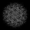 | |||||||||
|---|---|---|---|---|---|---|---|---|---|---|
| Title | A 3.3A sub-tomogram average of HIV-1 CA-SP1 from 5 tomograms in EMPIAR-10164 obtained using RELION 5 | |||||||||
 Map data Map data | Unmasked map after running the final 'Post-processing' job in RELION 5. | |||||||||
 Sample Sample |
| |||||||||
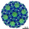
 Keywords Keywords | virus-like particle / enveloped / empty / VIRUS | |||||||||
| Biological species |   Human immunodeficiency virus 1 Human immunodeficiency virus 1 | |||||||||
| Method | subtomogram averaging / cryo EM / Resolution: 3.3 Å | |||||||||
 Authors Authors | Toader B / Scheres SHW | |||||||||
| Funding support |  United Kingdom, 1 items United Kingdom, 1 items
| |||||||||
 Citation Citation | Journal: FEBS Open Bio / Year: 2024 Title: An image processing pipeline for electron cryo-tomography in RELION-5. Authors: Alister Burt / Bogdan Toader / Rangana Warshamanage / Andriko von Kügelgen / Euan Pyle / Jasenko Zivanov / Dari Kimanius / Tanmay A M Bharat / Sjors H W Scheres /  Abstract: Electron tomography of frozen, hydrated samples allows structure determination of macromolecular complexes that are embedded in complex environments. Provided that the target complexes may be ...Electron tomography of frozen, hydrated samples allows structure determination of macromolecular complexes that are embedded in complex environments. Provided that the target complexes may be localised in noisy, three-dimensional tomographic reconstructions, averaging images of multiple instances of these molecules can lead to structures with sufficient resolution for de novo atomic modelling. Although many research groups have contributed image processing tools for these tasks, a lack of standardisation and interoperability represents a barrier for newcomers to the field. Here, we present an image processing pipeline for electron tomography data in RELION-5, with functionality ranging from the import of unprocessed movies to the automated building of atomic models in the final maps. Our explicit definition of metadata items that describe the steps of our pipeline has been designed for interoperability with other software tools and provides a framework for further standardisation. #1: Journal: Biorxiv / Year: 2024Title: An image processing pipeline for electron cryo-tomography in RELION-5 Authors: Burt A / Toader B / Warshamanage R / von Kugelgen A / Pyle E / Zivanov J / Kimanius D / Bharat T / Scheres S | |||||||||
| History |
|
- Structure visualization
| Supplemental images |
|---|

- Downloads & links
-EMDB archive
| Map data | emd_50672.map.gz | 24.2 MB |  EMDB map data format EMDB map data format | |
|---|---|---|---|---|
| Header (meta data) | emd-50672-v30.xmlemd-50672.xml | 15.1 KB 15.1 KB | Display Display | EMDB header |
| FSC (resolution estimation) | emd_50672_fsc.xml | 6.9 KB | Display | FSC data file |
| Images |  emd_50672.png emd_50672.png | 178.6 KB | ||
| Masks | emd_50672_msk_1.map | 27 MB | Mask map | |
| Filedesc metadata | emd-50672.cif.gz | 4.3 KB | ||
| Others | emd_50672_half_map_1.map.gzemd_50672_half_map_2.map.gz | 12.6 MB 12.6 MB | ||
| Archive directory |  http://ftp.pdbj.org/pub/emdb/structures/EMD-50672ftp://ftp.pdbj.org/pub/emdb/structures/EMD-50672 http://ftp.pdbj.org/pub/emdb/structures/EMD-50672ftp://ftp.pdbj.org/pub/emdb/structures/EMD-50672 | HTTPS FTP |
-Related structure data

-Links
| EMDB pages | EMDB (EBI/PDBe) / EMDataResource |
|---|---|
| Related items in Molecule of the Month |

-Map
| File | Download / File: emd_50672.map.gz / Format: CCP4 / Size: 27 MB / Type: IMAGE STORED AS FLOATING POINT NUMBER (4 BYTES) | ||||||||||||||||||||||||||||||||||||
|---|---|---|---|---|---|---|---|---|---|---|---|---|---|---|---|---|---|---|---|---|---|---|---|---|---|---|---|---|---|---|---|---|---|---|---|---|---|
| Annotation | Unmasked map after running the final 'Post-processing' job in RELION 5. | ||||||||||||||||||||||||||||||||||||
| Projections & slices | Image control
Images are generated by Spider. | ||||||||||||||||||||||||||||||||||||
| Voxel size | X=Y=Z: 1.35 Å | ||||||||||||||||||||||||||||||||||||
| Density |
| ||||||||||||||||||||||||||||||||||||
| Symmetry | Space group: 1 | ||||||||||||||||||||||||||||||||||||
| Details | EMDB XML:
|
 Z (Sec.)
 Y (Row.)
 X (Col.)


-Supplemental data
-Mask #1
| File | emd_50672_msk_1.map | ||||||||||||
|---|---|---|---|---|---|---|---|---|---|---|---|---|---|
| Projections & Slices |
| ||||||||||||
| Density Histograms |


-Half map: Half-map from the final 'Reconstruct particle' job in RELION 5.
| File | emd_50672_half_map_1.map | ||||||||||||
|---|---|---|---|---|---|---|---|---|---|---|---|---|---|
| Annotation | Half-map from the final 'Reconstruct particle' job in RELION 5. | ||||||||||||
| Projections & Slices |
| ||||||||||||
| Density Histograms |


-Half map: Half-map from the final 'Reconstruct particle' job in RELION 5.
| File | emd_50672_half_map_2.map | ||||||||||||
|---|---|---|---|---|---|---|---|---|---|---|---|---|---|
| Annotation | Half-map from the final 'Reconstruct particle' job in RELION 5. | ||||||||||||
| Projections & Slices |
| ||||||||||||
| Density Histograms |


- Sample components
-Entire : Human immunodeficiency virus 1
| Entire | Name: Human immunodeficiency virus 1 |
|---|---|
| Components |
|
-Supramolecule #1: Human immunodeficiency virus 1
| Supramolecule | Name: Human immunodeficiency virus 1 / type: virus / ID: 1 / Parent: 0 / NCBI-ID: 11676 / Sci species name: Human immunodeficiency virus 1 / Virus type: VIRUS-LIKE PARTICLE / Virus isolate: STRAIN / Virus enveloped: Yes / Virus empty: Yes |
|---|
-Experimental details
-Structure determination
| Method | cryo EM |
|---|---|
 Processing Processing | subtomogram averaging |
| Aggregation state | particle |
-Sample preparation
| Buffer | pH: 8 |
|---|---|
| Vitrification | Cryogen name: ETHANE |
- Electron microscopy
| Microscope | FEI TITAN KRIOS |
|---|---|
| Image recording | Film or detector model: GATAN K2 QUANTUM (4k x 4k) / Average electron dose: 3.0 e/Å2 |
| Electron beam | Acceleration voltage: 300 kV / Electron source:  FIELD EMISSION GUN FIELD EMISSION GUN |
| Electron optics | Illumination mode: OTHER / Imaging mode: BRIGHT FIELD / Nominal defocus max: 5.0 µm / Nominal defocus min: 1.5 µm |
| Experimental equipment |  Model: Titan Krios / Image courtesy: FEI Company |
-Image processing
| Final reconstruction | Applied symmetry - Point group: C6 (6 fold cyclic) / Resolution.type: BY AUTHOR / Resolution: 3.3 Å / Resolution method: FSC 0.143 CUT-OFF / Software - Name: RELION (ver. 5.0) / Number subtomograms used: 9038 |
|---|---|
| Extraction | Number tomograms: 5 / Number images used: 36088 / Software - Name: RELION (ver. 5.0) |
| Final angle assignment | Type: MAXIMUM LIKELIHOOD / Software - Name: RELION (ver. 5.0) |
| FSC plot (resolution estimation) |  |
