Movie
Movie Controller

[English] 日本語
 Yorodumi
- EMDB-50668: Cryo-EM structure of MmCAT1 bound with FrMLV-RBD in the apo inwar... -
+ Open data

- Basic information
| Entry |  | |||||||||
|---|---|---|---|---|---|---|---|---|---|---|
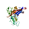
| Title | Cryo-EM structure of MmCAT1 bound with FrMLV-RBD in the apo inward-open state | |||||||||
 Map data Map data | main | |||||||||
 Sample Sample |
| |||||||||
 Keywords Keywords | MmCAT1 / FrMLV-RBD / SLC / viral tropism / MEMBRANE PROTEIN | |||||||||
| Function / homology |  Function and homology information Function and homology informationL-ornithine transmembrane transporter activity / L-lysine transmembrane transporter activity / lysine transport / L-ornithine transmembrane transport / L-lysine transmembrane transport / L-amino acid transport / L-histidine import across plasma membrane / ornithine transport / L-arginine import across plasma membrane / Amino acid transport across the plasma membrane ...L-ornithine transmembrane transporter activity / L-lysine transmembrane transporter activity / lysine transport / L-ornithine transmembrane transport / L-lysine transmembrane transport / L-amino acid transport / L-histidine import across plasma membrane / ornithine transport / L-arginine import across plasma membrane / Amino acid transport across the plasma membrane / basic amino acid transmembrane transporter activity / L-arginine transmembrane transport / L-histidine transmembrane transporter activity / L-arginine transmembrane transporter activity / amino acid import across plasma membrane / regulation of TOR signaling / amino acid transmembrane transporter activity / positive regulation of T cell proliferation / basal plasma membrane / bioluminescence / generation of precursor metabolites and energy / virus receptor activity / basolateral plasma membrane / apical plasma membrane / fusion of virus membrane with host plasma membrane / viral envelope / symbiont entry into host cell / virion attachment to host cell / host cell plasma membrane / virion membrane / protein-containing complex / membrane / metal ion binding / plasma membrane Similarity search - Function | |||||||||
| Biological species |   Murine leukemia virus Murine leukemia virus | |||||||||
| Method | single particle reconstruction / cryo EM / Resolution: 3.5 Å | |||||||||
 Authors Authors | Ye M / Zhou D / Pike ACW / Wang S / Wang D / Bakshi S / Brooke L / Williams E / Elkins J / Stuart DI / Sauer DB | |||||||||
| Funding support | 1 items
| |||||||||
 Citation Citation | Journal: Nat Commun / Year: 2026 Title: Amino acid and viral binding by the high-affinity Cationic Amino acid Transporter 1 (CAT1) from Mus musculus. Authors: Mingda Ye / Zhu Liang / Daming Zhou / Ashley C W Pike / SiYi Wang / Dong Wang / Souvika Bakshi / Laurent Brooke / Eleanor P Williams / Jonathan M Elkins / Benedikt M Kessler / David I Stuart / David B Sauer /   Abstract: Arginine, lysine, and ornithine are critical to several fundamental aspects of organismal physiology, including protein structure and function, the urea cycle, and intracellular signaling. These ...Arginine, lysine, and ornithine are critical to several fundamental aspects of organismal physiology, including protein structure and function, the urea cycle, and intracellular signaling. These cationic amino acids are imported by several membrane transporters, most notably the Cationic Amino acid Transporters (CATs) in the SLC7 family. Of these, CAT1 is also the receptor for two orthoretroviruses, and determines the host tropism for these viruses. Here, using a combination of CryoEM and in vitro biochemical techniques, we characterize the substrate recognition and transport of CAT1 from Mus musculus. Further, by determining the structures of MmCAT1 in complex with the receptor binding domain from the Friend Murine Leukemia Virus, we identify the key structural interactions that determine the virus' rodent-specific tropism. | |||||||||
| History |
|
- Structure visualization
| Supplemental images |
|---|

- Downloads & links
-EMDB archive
| Map data | emd_50668.map.gz | 122.2 MB | EMDB map data format | |
|---|---|---|---|---|
| Header (meta data) | emd-50668-v30.xmlemd-50668.xml | 25.7 KB 25.7 KB | Display Display | EMDB header |
| FSC (resolution estimation) | emd_50668_fsc.xml | 10.7 KB | Display | FSC data file |
| Images |  emd_50668.png emd_50668.png | 57 KB | ||
| Masks | emd_50668_msk_1.map | 129.7 MB | Mask map | |
| Filedesc metadata | emd-50668.cif.gz | 7.8 KB | ||
| Others | emd_50668_half_map_1.map.gzemd_50668_half_map_2.map.gz | 120.5 MB 120.5 MB | ||
| Archive directory |  http://ftp.pdbj.org/pub/emdb/structures/EMD-50668ftp://ftp.pdbj.org/pub/emdb/structures/EMD-50668 http://ftp.pdbj.org/pub/emdb/structures/EMD-50668ftp://ftp.pdbj.org/pub/emdb/structures/EMD-50668 | HTTPS FTP |
-Related structure data
| Related structure data |  9fqtMC  9fquC  9fqvC  9fqwC M: atomic model generated by this map C: citing same article ( |
|---|---|
| Similar structure data |


-Links
| EMDB pages | EMDB (EBI/PDBe) / EMDataResource |
|---|---|
| Related items in Molecule of the Month |


-Map
| File | Download / File: emd_50668.map.gz / Format: CCP4 / Size: 129.7 MB / Type: IMAGE STORED AS FLOATING POINT NUMBER (4 BYTES) | ||||||||||||||||||||||||||||||||||||
|---|---|---|---|---|---|---|---|---|---|---|---|---|---|---|---|---|---|---|---|---|---|---|---|---|---|---|---|---|---|---|---|---|---|---|---|---|---|
| Annotation | main | ||||||||||||||||||||||||||||||||||||
| Projections & slices | Image control
Images are generated by Spider. | ||||||||||||||||||||||||||||||||||||
| Voxel size | X=Y=Z: 0.825 Å | ||||||||||||||||||||||||||||||||||||
| Density |
| ||||||||||||||||||||||||||||||||||||
| Symmetry | Space group: 1 | ||||||||||||||||||||||||||||||||||||
| Details | EMDB XML:
|
 Z (Sec.)
 Y (Row.)
 X (Col.)


-Supplemental data
-Mask #1
| File | emd_50668_msk_1.map | ||||||||||||
|---|---|---|---|---|---|---|---|---|---|---|---|---|---|
| Projections & Slices |
| ||||||||||||
| Density Histograms |


-Half map: half A
| File | emd_50668_half_map_1.map | ||||||||||||
|---|---|---|---|---|---|---|---|---|---|---|---|---|---|
| Annotation | half A | ||||||||||||
| Projections & Slices |
| ||||||||||||
| Density Histograms |


-Half map: half B
| File | emd_50668_half_map_2.map | ||||||||||||
|---|---|---|---|---|---|---|---|---|---|---|---|---|---|
| Annotation | half B | ||||||||||||
| Projections & Slices |
| ||||||||||||
| Density Histograms |


- Sample components
-Entire : MmCAT1(apo) bound with FrMLV-RBD
| Entire | Name: MmCAT1(apo) bound with FrMLV-RBD |
|---|---|
| Components |
|
-Supramolecule #1: MmCAT1(apo) bound with FrMLV-RBD
| Supramolecule | Name: MmCAT1(apo) bound with FrMLV-RBD / type: complex / ID: 1 / Parent: 0 / Macromolecule list: #1 |
|---|---|
| Source (natural) | Organism: |
| Molecular weight | Theoretical: 128.8 KDa |
-Macromolecule #1: High affinity cationic amino acid transporter 1,Green fluorescent...
| Macromolecule | Name: High affinity cationic amino acid transporter 1,Green fluorescent protein type: protein_or_peptide / ID: 1 / Number of copies: 1 / Enantiomer: LEVO |
|---|---|
| Source (natural) | Organism: |
| Molecular weight | Theoretical: 98.703758 KDa |
| Recombinant expression | Organism:  Homo sapiens (human) Homo sapiens (human) |
| Sequence | String: MLRRKVVDCS REESRLSRCL NTYDLVALGV GSTLGAGVYV LAGAVARENA GPAIVISFLI AALASVLAGL CYGEFGARVP KTGSAYLYS YVTVGELWAF ITGWNLILSY IIGTSSVARA WSATFDELIG KPIGEFSRQH MALNAPGVLA QTPDIFAVII I IILTGLLT ...String: MLRRKVVDCS REESRLSRCL NTYDLVALGV GSTLGAGVYV LAGAVARENA GPAIVISFLI AALASVLAGL CYGEFGARVP KTGSAYLYS YVTVGELWAF ITGWNLILSY IIGTSSVARA WSATFDELIG KPIGEFSRQH MALNAPGVLA QTPDIFAVII I IILTGLLT LGVKESAMVN KIFTCINVLV LCFIVVSGFV KGSIKNWQLT EKNFSCNNND TNVKYGEGGF MPFGFSGVLS GA ATCFYAF VGFDCIATTG EEVKNPQKAI PVGIVASLLI CFIAYFGVSA ALTLMMPYFC LDIDSPLPGA FKHQGWEEAK YAV AIGSLC ALSTSLLGSM FPMPRVIYAM AEDGLLFKFL AKINNRTKTP VIATVTSGAI AAVMAFLFEL KDLVDLMSIG TLLA YSLVA ACVLVLRYQP EQPNLVYQMA RTTEELDRVD QNELVSASES QTGFLPVAEK FSLKSILSPK NVEPSKFSGL IVNIS AGLL AALIITVCIV AVLGREALAE GTLWAVFVMT GSVLLCMLVT GIIWRQPESK TKLSFKVPFV PVLPVLSIFV NIYLMM QLD QGTWVRFAVW MLIGFTIYFG YGIWHSEEAS LAAGQAKTPD SNLDQCKAEN LYFQSGSAVS KGEELFTGVV PILVELD GD VNGHKFSVSG EGEGDATYGK LTLKFICTTG KLPVPWPTLV TTLTYGVQCF SRYPDHMKQH DFFKSAMPEG YVQERTIF F KDDGNYKTRA EVKFEGDTLV NRIELKGIDF KEDGNILGHK LEYNYNSHNV YIMADKQKNG IKVNFKIRHN IEDGSVQLA DHYQQNTPIG DGPVLLPDNH YLSTQSKLSK DPNEKRDHMV LLEFVTAAGI TLGMDELYKS GLRSWSHPQF EKGGGSGGGS GGSAWSHPQ FEKHHHHHHH HHH UniProtKB: High affinity cationic amino acid transporter 1, Green fluorescent protein |
-Macromolecule #2: Surface protein
| Macromolecule | Name: Surface protein / type: protein_or_peptide / ID: 2 / Number of copies: 1 / Enantiomer: LEVO |
|---|---|
| Source (natural) | Organism: Murine leukemia virus |
| Molecular weight | Theoretical: 30.272078 KDa |
| Recombinant expression | Organism: Homo sapiens (human) |
| Sequence | String: MGILPSPGMP ALLSLVSLLS VLLMGCVAET GAAPGSSPHQ VYNITWEVTN GDRETVWAIS GNHPLWTWWP VLTPDLCMLA LSGPPHWGL EYQAPYSSPP GPPCCSGSSG SSAGCSRDCD EPLTSLTPRC NTAWNRLKLD QVTHKSSEGF YVCPGSHRPR E AKSCGGPD ...String: MGILPSPGMP ALLSLVSLLS VLLMGCVAET GAAPGSSPHQ VYNITWEVTN GDRETVWAIS GNHPLWTWWP VLTPDLCMLA LSGPPHWGL EYQAPYSSPP GPPCCSGSSG SSAGCSRDCD EPLTSLTPRC NTAWNRLKLD QVTHKSSEGF YVCPGSHRPR E AKSCGGPD SFYCASWGCE TTGRVYWKPS SSWDYITVDN NLTTSQAVQV CKDNKWCNPL AIQFTNAGKQ VTSWTTGHYW GL RLYVSGR DPGLTFGIRL RYQNLGPRVP GTKHHHHHH UniProtKB: Envelope glycoprotein |
-Macromolecule #5: 2-acetamido-2-deoxy-beta-D-glucopyranose
| Macromolecule | Name: 2-acetamido-2-deoxy-beta-D-glucopyranose / type: ligand / ID: 5 / Number of copies: 1 / Formula: NAG |
|---|---|
| Molecular weight | Theoretical: 221.208 Da |
| Chemical component information |  ChemComp-NAG: |
-Macromolecule #6: CHOLESTEROL HEMISUCCINATE
| Macromolecule | Name: CHOLESTEROL HEMISUCCINATE / type: ligand / ID: 6 / Number of copies: 1 / Formula: Y01 |
|---|---|
| Molecular weight | Theoretical: 486.726 Da |
| Chemical component information |  ChemComp-Y01: |
-Experimental details
-Structure determination
| Method | cryo EM |
|---|---|
 Processing Processing | single particle reconstruction |
| Aggregation state | particle |
-Sample preparation
| Concentration | 3.8 mg/mL | |||||||||||||||
|---|---|---|---|---|---|---|---|---|---|---|---|---|---|---|---|---|
| Buffer | pH: 7.5 Component:
| |||||||||||||||
| Grid | Model: Quantifoil R1.2/1.3 / Pretreatment - Type: GLOW DISCHARGE | |||||||||||||||
| Vitrification | Cryogen name: ETHANE |
- Electron microscopy
| Microscope | FEI TITAN KRIOS |
|---|---|
| Image recording | Film or detector model: GATAN K3 (6k x 4k) / Average electron dose: 50.0 e/Å2 |
| Electron beam | Acceleration voltage: 300 kV / Electron source:  FIELD EMISSION GUN FIELD EMISSION GUN |
| Electron optics | Illumination mode: FLOOD BEAM / Imaging mode: BRIGHT FIELD / Nominal defocus max: 2.6 µm / Nominal defocus min: 1.2 µm |
| Experimental equipment |  Model: Titan Krios / Image courtesy: FEI Company |