National Institutes of Health/National Institute on Aging (NIH/NIA)
P30-AG010133
米国
National Institutes of Health/National Institute on Aging (NIH/NIA)
P30-AG072976
米国
National Institutes of Health/National Institute on Aging (NIH/NIA)
R01-NS110437
米国
引用
ジャーナル: Acta Neuropathol / 年: 2024 タイトル: TMEM106B amyloid filaments in the Biondi bodies of ependymal cells. 著者: Bernardino Ghetti / Manuel Schweighauser / Max H Jacobsen / Derrick Gray / Mehtap Bacioglu / Alexey G Murzin / Bradley S Glazier / Taxiarchis Katsinelos / Ruben Vidal / Kathy L Newell / ...著者: Bernardino Ghetti / Manuel Schweighauser / Max H Jacobsen / Derrick Gray / Mehtap Bacioglu / Alexey G Murzin / Bradley S Glazier / Taxiarchis Katsinelos / Ruben Vidal / Kathy L Newell / Sujuan Gao / Holly J Garringer / Maria Grazia Spillantini / Sjors H W Scheres / Michel Goedert / 要旨: Biondi bodies are filamentous amyloid inclusions of unknown composition in ependymal cells of the choroid plexuses, ependymal cells lining cerebral ventricles and ependymal cells of the central canal ...Biondi bodies are filamentous amyloid inclusions of unknown composition in ependymal cells of the choroid plexuses, ependymal cells lining cerebral ventricles and ependymal cells of the central canal of the spinal cord. Their formation is age-dependent and they are commonly associated with a variety of neurodegenerative conditions, including Alzheimer's disease and Lewy body disorders. Here, we show that Biondi bodies are strongly immunoreactive with TMEM239, an antibody specific for inclusions of transmembrane protein 106B (TMEM106B). Biondi bodies were labelled by both this antibody and the amyloid dye pFTAA. Many Biondi bodies were also labelled for TMEM106B and the lysosomal markers Hexosaminidase A and Cathepsin D. By transmission immuno-electron microscopy, Biondi bodies of choroid plexuses were decorated by TMEM239 and were associated with structures that resembled residual bodies or secondary lysosomes. By electron cryo-microscopy, TMEM106B filaments from Biondi bodies of choroid plexuses were similar (Biondi variant), but not identical, to the fold I that was previously identified in filaments from brain parenchyma.
 ムービー
ムービー コントローラー


 データを開く
 基本情報
基本情報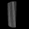
 マップデータ
 試料
試料 キーワード
キーワード 機能・相同性情報
 Homo sapiens (ヒト)
 データ登録者
 英国,
英国,  米国, 5件
  引用
引用 構造の表示
構造の表示
 ダウンロードとリンク
 emd_50587.png
 http://ftp.pdbj.org/pub/emdb/structures/EMD-50587
 Z (Sec.)
 Y (Row.)
 X (Col.)


 試料の構成要素
 解析
解析 電子顕微鏡法
 FIELD EMISSION GUN