Movie
Movie Controller


+ Open data

- Basic information
| Entry | Database: PDB / ID: 9fnb | ||||||||||||||||||
|---|---|---|---|---|---|---|---|---|---|---|---|---|---|---|---|---|---|---|---|
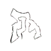
| Title | TMEM106B filaments from Biondi bodies (Biondi variant) | ||||||||||||||||||
 Components Components | Transmembrane protein 106B | ||||||||||||||||||
 Keywords Keywords | PROTEIN FIBRIL / TMEM106B / Amyloid / Biondi bodies / Choroid plexus | ||||||||||||||||||
| Function / homology |  Function and homology information Function and homology informationlysosomal protein catabolic process / lysosomal lumen acidification / lysosome localization / regulation of lysosome organization / positive regulation of dendrite development / lysosomal transport / dendrite morphogenesis / lysosome organization / neuron cellular homeostasis / late endosome membrane ...lysosomal protein catabolic process / lysosomal lumen acidification / lysosome localization / regulation of lysosome organization / positive regulation of dendrite development / lysosomal transport / dendrite morphogenesis / lysosome organization / neuron cellular homeostasis / late endosome membrane / ATPase binding / lysosome / endosome / lysosomal membrane / plasma membrane Similarity search - Function | ||||||||||||||||||
| Biological species |  Homo sapiens (human) Homo sapiens (human) | ||||||||||||||||||
| Method | ELECTRON MICROSCOPY / helical reconstruction / cryo EM / Resolution: 2.64 Å | ||||||||||||||||||
 Authors Authors | Ghetti, B. / Schweighauser, M. / Jacobsen, M.H. / Gray, D. / Bacioglu, M. / Murzin, A.G. / Glazier, B.S. / Vidal, R. / Newell, K.L. / Gao, S. ...Ghetti, B. / Schweighauser, M. / Jacobsen, M.H. / Gray, D. / Bacioglu, M. / Murzin, A.G. / Glazier, B.S. / Vidal, R. / Newell, K.L. / Gao, S. / Garringer, H.J. / Spillantini, M.G. / Scheres, S.H.W. / Goedert, M. | ||||||||||||||||||
| Funding support |  United Kingdom, United Kingdom,  United States, 5items United States, 5items
| ||||||||||||||||||
 Citation Citation | Journal: Acta Neuropathol / Year: 2024 Title: TMEM106B amyloid filaments in the Biondi bodies of ependymal cells. Authors: Bernardino Ghetti / Manuel Schweighauser / Max H Jacobsen / Derrick Gray / Mehtap Bacioglu / Alexey G Murzin / Bradley S Glazier / Taxiarchis Katsinelos / Ruben Vidal / Kathy L Newell / ...Authors: Bernardino Ghetti / Manuel Schweighauser / Max H Jacobsen / Derrick Gray / Mehtap Bacioglu / Alexey G Murzin / Bradley S Glazier / Taxiarchis Katsinelos / Ruben Vidal / Kathy L Newell / Sujuan Gao / Holly J Garringer / Maria Grazia Spillantini / Sjors H W Scheres / Michel Goedert / Abstract: Biondi bodies are filamentous amyloid inclusions of unknown composition in ependymal cells of the choroid plexuses, ependymal cells lining cerebral ventricles and ependymal cells of the central canal ...Biondi bodies are filamentous amyloid inclusions of unknown composition in ependymal cells of the choroid plexuses, ependymal cells lining cerebral ventricles and ependymal cells of the central canal of the spinal cord. Their formation is age-dependent and they are commonly associated with a variety of neurodegenerative conditions, including Alzheimer's disease and Lewy body disorders. Here, we show that Biondi bodies are strongly immunoreactive with TMEM239, an antibody specific for inclusions of transmembrane protein 106B (TMEM106B). Biondi bodies were labelled by both this antibody and the amyloid dye pFTAA. Many Biondi bodies were also labelled for TMEM106B and the lysosomal markers Hexosaminidase A and Cathepsin D. By transmission immuno-electron microscopy, Biondi bodies of choroid plexuses were decorated by TMEM239 and were associated with structures that resembled residual bodies or secondary lysosomes. By electron cryo-microscopy, TMEM106B filaments from Biondi bodies of choroid plexuses were similar (Biondi variant), but not identical, to the fold I that was previously identified in filaments from brain parenchyma. | ||||||||||||||||||
| History |
|
- Structure visualization
| Structure viewer | Molecule: MolmilJmol/JSmol |
|---|
- Downloads & links
-Download
| PDBx/mmCIF format | 9fnb.cif.gz | 120 KB | Display | PDBx/mmCIF format |
|---|---|---|---|---|
| PDB format | pdb9fnb.ent.gz | 95.5 KB | Display | PDB format |
| PDBx/mmJSON format | 9fnb.json.gz | Tree view | PDBx/mmJSON format | |
| Others |  Other downloads Other downloads |
-Validation report
| Arichive directory | https://data.pdbj.org/pub/pdb/validation_reports/fn/9fnbftp://data.pdbj.org/pub/pdb/validation_reports/fn/9fnb | HTTPS FTP |
|---|
-Related structure data
| Related structure data | 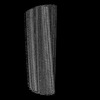 50587MC M: map data used to model this data C: citing same article ( |
|---|---|
| Similar structure data |
-Links
 PDBj
PDBj
- Assembly
| Deposited unit | 
|
|---|---|
| 1 |
|
-Components
| #1: Protein | Mass: 15502.680 Da / Num. of mol.: 4 / Source method: isolated from a natural source / Source: (natural) Homo sapiens (human) / References: UniProt: Q9NUM4#2: Sugar | ChemComp-NAG /   Type: D-saccharide, beta linking / Mass: 221.208 Da / Num. of mol.: 16 / Source method: obtained synthetically / Formula: C8H15NO6 Type: D-saccharide, beta linking / Mass: 221.208 Da / Num. of mol.: 16 / Source method: obtained synthetically / Formula: C8H15NO6Has ligand of interest | N | Has protein modification | Y | |
|---|
-Experimental details
-Experiment
| Experiment | Method: ELECTRON MICROSCOPY |
|---|---|
| EM experiment | Aggregation state: FILAMENT / 3D reconstruction method: helical reconstruction |
- Sample preparation
| Component | Name: TMEM106B Filaments / Type: TISSUE Details: TMEM106B filaments extracted from Biondi bodies of Alzheimer's disease Entity ID: #1 / Source: NATURAL |
|---|---|
| Source (natural) | Organism: Homo sapiens (human) / Organ: Brain / Tissue: Choroid plexus |
| Buffer solution | pH: 7.4 |
| Specimen | Embedding applied: NO / Shadowing applied: NO / Staining applied: NO / Vitrification applied: YES |
| Vitrification | Instrument: FEI VITROBOT MARK IV / Cryogen name: ETHANE / Humidity: 100 % / Chamber temperature: 277 K |
- Electron microscopy imaging
| Experimental equipment |  Model: Titan Krios / Image courtesy: FEI Company |
|---|---|
| Microscopy | Model: FEI TITAN KRIOS |
| Electron gun | Electron source:  FIELD EMISSION GUN / Accelerating voltage: 300 kV / Illumination mode: FLOOD BEAM FIELD EMISSION GUN / Accelerating voltage: 300 kV / Illumination mode: FLOOD BEAM |
| Electron lens | Mode: BRIGHT FIELD / Nominal magnification: 96000 X / Nominal defocus max: 2400 nm / Nominal defocus min: 1700 nm / Alignment procedure: COMA FREE |
| Specimen holder | Cryogen: NITROGEN |
| Image recording | Electron dose: 30 e/Å2 / Film or detector model: FEI FALCON IV (4k x 4k) |
- Processing
| EM software |
| ||||||||||||||||||||||||
|---|---|---|---|---|---|---|---|---|---|---|---|---|---|---|---|---|---|---|---|---|---|---|---|---|---|
| CTF correction | Type: PHASE FLIPPING AND AMPLITUDE CORRECTION | ||||||||||||||||||||||||
| Helical symmerty | Angular rotation/subunit: -0.46 ° / Axial rise/subunit: 4.88 Å / Axial symmetry: C1 | ||||||||||||||||||||||||
| Particle selection | Num. of particles selected: 685561 | ||||||||||||||||||||||||
| 3D reconstruction | Resolution: 2.64 Å / Resolution method: FSC 0.143 CUT-OFF / Num. of particles: 38763 / Symmetry type: HELICAL | ||||||||||||||||||||||||
| Atomic model building | PDB-ID: 7QVC Accession code: 7QVC / Source name: PDB / Type: experimental model |