Journal: Curr Biol / Year: 2024 Title: Protofilament-specific nanopatterns of tubulin post-translational modifications regulate the mechanics of ciliary beating. Authors: Gonzalo Alvarez Viar / Nikolai Klena / Fabrizio Martino / Adrian Pascal Nievergelt / Davide Bolognini / Paola Capasso / Gaia Pigino / Abstract: Controlling ciliary beating is essential for motility and signaling in eukaryotes. This process relies on the regulation of various axonemal proteins that assemble in stereotyped patterns onto ...Controlling ciliary beating is essential for motility and signaling in eukaryotes. This process relies on the regulation of various axonemal proteins that assemble in stereotyped patterns onto individual microtubules of the ciliary structure. Additionally, each axonemal protein interacts exclusively with determined tubulin protofilaments of the neighboring microtubule to carry out its function. While it is known that tubulin post-translational modifications (PTMs) are important for proper ciliary motility, the mode and extent to which they contribute to these interactions remain poorly understood. Currently, the prevailing understanding is that PTMs can confer functional specialization at the level of individual microtubules. However, this paradigm falls short of explaining how the tubulin code can manage the complexity of the axonemal structure where functional interactions happen in defined patterns at the sub-microtubular scale. Here, we combine immuno-cryo-electron tomography (cryo-ET), expansion microscopy, and mutant analysis to show that, in motile cilia, tubulin glycylation and polyglutamylation form mutually exclusive protofilament-specific nanopatterns at a sub-microtubular scale. These nanopatterns are consistent with the distributions of axonemal dyneins and nexin-dynein regulatory complexes, respectively, and are indispensable for their regulation during ciliary beating. Our findings offer a new paradigm for understanding how different tubulin PTMs, such as glycylation, glutamylation, acetylation, tyrosination, and detyrosination, can coexist within the ciliary structure and specialize individual protofilaments for the regulation of diverse protein complexes. The identification of a ciliary tubulin nanocode by cryo-ET suggests the need for high-resolution studies to better understand the molecular role of PTMs in other cellular compartments beyond the cilium.
In the structure databanks used in Yorodumi, some data are registered as the other names, "COVID-19 virus" and "2019-nCoV". Here are the details of the virus and the list of structure data.
Jan 31, 2019. EMDB accession codes are about to change! (news from PDBe EMDB page)
EMDB accession codes are about to change! (news from PDBe EMDB page)
The allocation of 4 digits for EMDB accession codes will soon come to an end. Whilst these codes will remain in use, new EMDB accession codes will include an additional digit and will expand incrementally as the available range of codes is exhausted. The current 4-digit format prefixed with “EMD-” (i.e. EMD-XXXX) will advance to a 5-digit format (i.e. EMD-XXXXX), and so on. It is currently estimated that the 4-digit codes will be depleted around Spring 2019, at which point the 5-digit format will come into force.
The EM Navigator/Yorodumi systems omit the EMD- prefix.
Related info.:Q: What is EMD? / ID/Accession-code notation in Yorodumi/EM Navigator
Yorodumi is a browser for structure data from EMDB, PDB, SASBDB, etc.
This page is also the successor to EM Navigator detail page, and also detail information page/front-end page for Omokage search.
The word "yorodu" (or yorozu) is an old Japanese word meaning "ten thousand". "mi" (miru) is to see.
Related info.:EMDB / PDB / SASBDB / Comparison of 3 databanks / Yorodumi Search / Aug 31, 2016. New EM Navigator & Yorodumi / Yorodumi Papers / Jmol/JSmol / Function and homology information / Changes in new EM Navigator and Yorodumi
 Movie
Movie Controller

 Yorodumi
 Open data
 Basic information
 Map data
 Sample
 Keywords
 C.reinhardtii (plant)
 Authors
 Citation

 Structure visualization
 Downloads & links
 EMDB map data format
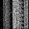
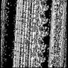
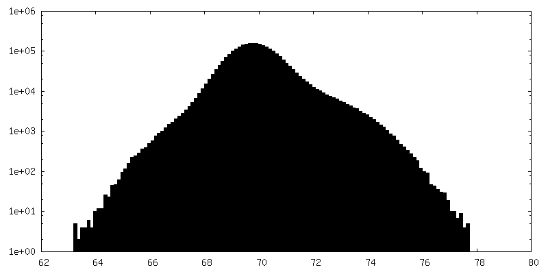
 emd_50565.png
 http://ftp.pdbj.org/pub/emdb/structures/EMD-50565


 Z (Sec.)
 Y (Row.)
 X (Col.)


 Sample components
 Processing
 Electron microscopy
 FIELD EMISSION GUN