- EMDB-50516: Structure of the DNase I- and phalloidin-bound pointed end of F-a... -
+
Open data
ID or keywords:
Loading...
-
Basic information
Entry
Database: EMDB / ID: EMD-50516
Title
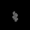
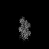
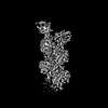
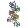
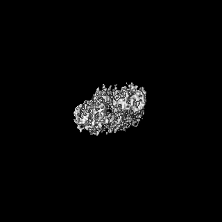
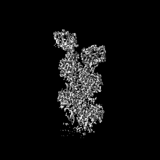
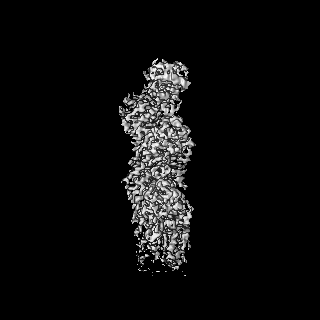
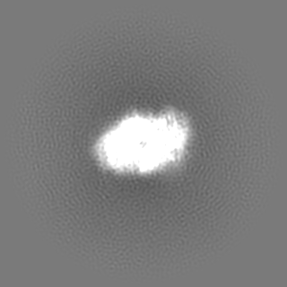
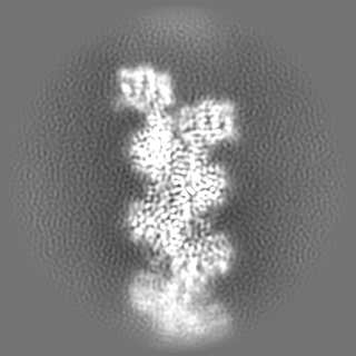
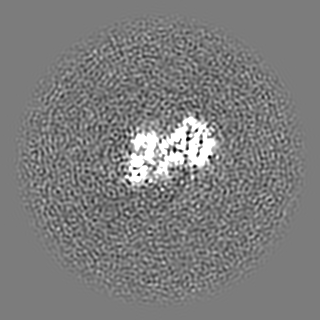
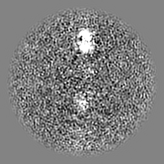
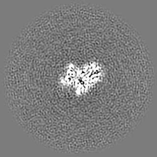
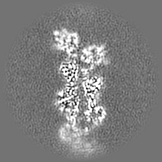
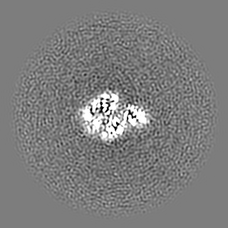
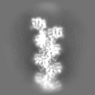
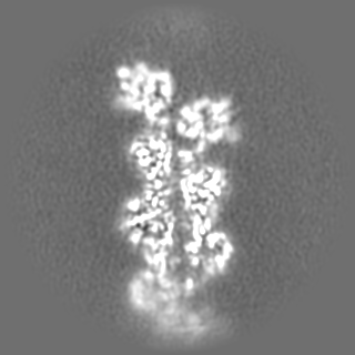
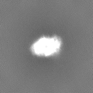
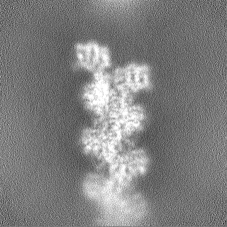
Structure of the DNase I- and phalloidin-bound pointed end of F-actin (conformer 1)
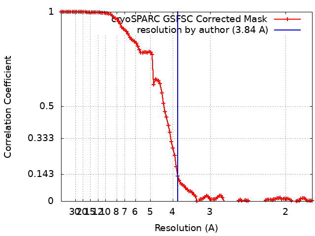
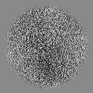
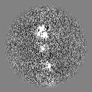
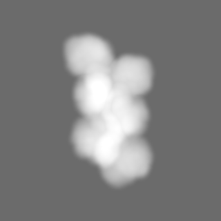
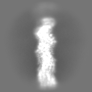
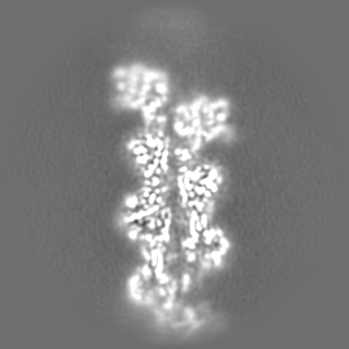
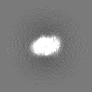
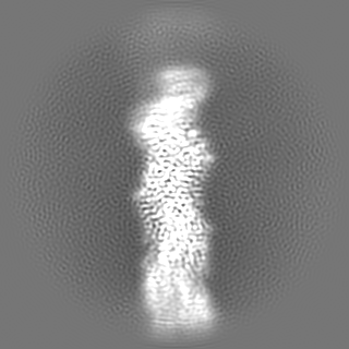
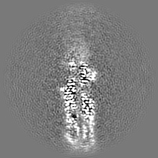
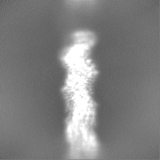
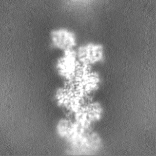
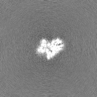
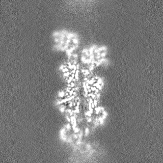
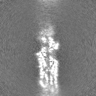
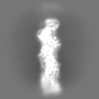
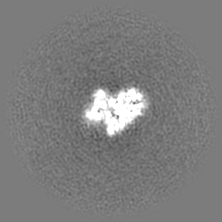
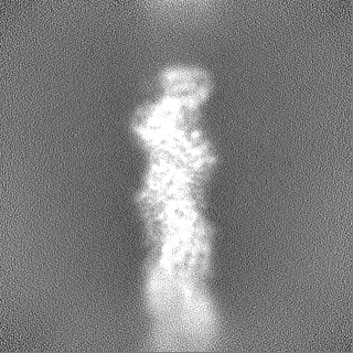
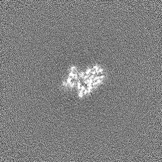
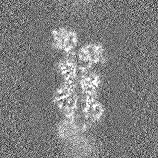
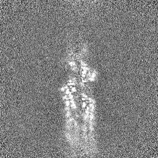
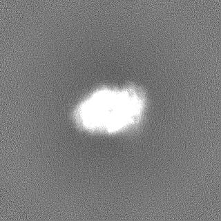
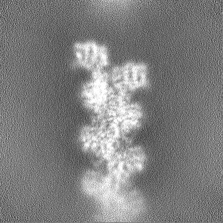
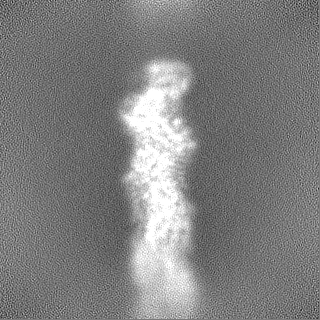
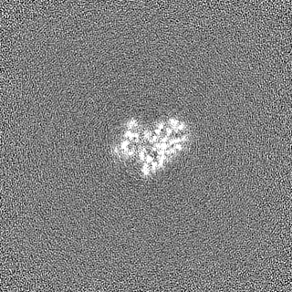
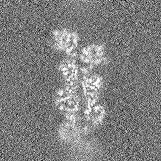
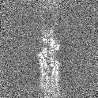
Map data
Sharpened cryo-EM density map of the DNase I- and phalloidin-bound pointed end of actin filaments (conformer 1).
Sample
Complex: F-actin pointed end bound by DNase I and phalloidin.
Complex: Actin filament pointed end
Protein or peptide: Actin, cytoplasmic 1, N-terminally processed
Complex: Phalloidin
Protein or peptide: Phalloidin
Complex: Two DNase I molecules, each bound to the ultimate and penultimate actin subunits of the F-actin pointed end.
Protein or peptide: Deoxyribonuclease-1
Ligand: ADENOSINE-5'-DIPHOSPHATE
Ligand: MAGNESIUM ION
Ligand: PHOSPHATE ION
Keywords
actin / phalloidin / filament / pointed end / DNase I / STRUCTURAL PROTEIN
Function / homology
Function and homology information
Cell-extracellular matrix interactions / Formation of the canonical BAF (cBAF) complex / Formation of the polybromo-BAF (pBAF) complex / Formation of the embryonic stem cell BAF (esBAF) complex / Formation of the non-canonical BAF (ncBAF) complex / Formation of neuronal progenitor and neuronal BAF (npBAF and nBAF) / Adherens junctions interactions / Regulation of CDH1 Function / Formation of the dystrophin-glycoprotein complex (DGC) / regulation of neutrophil mediated cytotoxicity ...Cell-extracellular matrix interactions / Formation of the canonical BAF (cBAF) complex / Formation of the polybromo-BAF (pBAF) complex / Formation of the embryonic stem cell BAF (esBAF) complex / Formation of the non-canonical BAF (ncBAF) complex / Formation of neuronal progenitor and neuronal BAF (npBAF and nBAF) / Adherens junctions interactions / Regulation of CDH1 Function / Formation of the dystrophin-glycoprotein complex (DGC) / regulation of neutrophil mediated cytotoxicity / B-WICH complex positively regulates rRNA expression / zymogen granule / RHO GTPases activate IQGAPs / Gap junction degradation / Formation of annular gap junctions / RHO GTPases Activate Formins / regulation of acute inflammatory response / RHOF GTPase cycle / EPHB-mediated forward signaling / Regulation of actin dynamics for phagocytic cup formation / RHO GTPases Activate WASPs and WAVEs / MAP2K and MAPK activation / DNA Damage Recognition in GG-NER / deoxyribonuclease I / UCH proteinases / neutrophil activation involved in immune response / VEGFA-VEGFR2 Pathway / deoxyribonuclease I activity / cellular response to cytochalasin B / Clathrin-mediated endocytosis / regulation of transepithelial transport / morphogenesis of a polarized epithelium / structural constituent of postsynaptic actin cytoskeleton / protein localization to adherens junction / dense body / Tat protein binding / postsynaptic actin cytoskeleton / apical protein localization / adherens junction assembly / DNA catabolic process / tight junction / apical junction complex / regulation of norepinephrine uptake / transporter regulator activity / NuA4 histone acetyltransferase complex / cortical cytoskeleton / establishment or maintenance of cell polarity / nitric-oxide synthase binding / brush border / regulation of synaptic vesicle endocytosis / kinesin binding / regulation of protein localization to plasma membrane / positive regulation of double-strand break repair via homologous recombination / axonogenesis / calyx of Held / nitric-oxide synthase regulator activity / cell motility / actin filament / adherens junction / Hydrolases; Acting on acid anhydrides; Acting on acid anhydrides to facilitate cellular and subcellular movement / Schaffer collateral - CA1 synapse / cytoplasmic ribonucleoprotein granule / actin cytoskeleton / nuclear envelope / nucleosome / lamellipodium / actin binding / cytoskeleton / regulation of cell cycle / ribonucleoprotein complex / axon / focal adhesion / apoptotic process / synapse / protein kinase binding / glutamatergic synapse / ATP hydrolysis activity / protein-containing complex / DNA binding / extracellular region / ATP binding / membrane / identical protein binding / nucleus / plasma membrane / cytosol / cytoplasm Similarity search - Function
Deoxyribonuclease I / Deoxyribonuclease I, active site / Deoxyribonuclease I, conservied site / Deoxyribonuclease I signature 2. / Deoxyribonuclease I signature 1. / deoxyribonuclease I / Endonuclease/exonuclease/phosphatase / Endonuclease/Exonuclease/phosphatase family / Endonuclease/exonuclease/phosphatase superfamily / Actins signature 1. ...Deoxyribonuclease I / Deoxyribonuclease I, active site / Deoxyribonuclease I, conservied site / Deoxyribonuclease I signature 2. / Deoxyribonuclease I signature 1. / deoxyribonuclease I / Endonuclease/exonuclease/phosphatase / Endonuclease/Exonuclease/phosphatase family / Endonuclease/exonuclease/phosphatase superfamily / Actins signature 1. / Actin, conserved site / Actins signature 2. / Actin/actin-like conserved site / Actins and actin-related proteins signature. / Actin / Actin family / Actin / ATPase, nucleotide binding domain Similarity search - Domain/homology
Journal: Nat Commun / Year: 2024 Title: Phalloidin and DNase I-bound F-actin pointed end structures reveal principles of filament stabilization and disassembly. Authors: Micaela Boiero Sanders / Wout Oosterheert / Oliver Hofnagel / Peter Bieling / Stefan Raunser / Abstract: Actin filament turnover involves subunits binding to and dissociating from the filament ends, with the pointed end being the primary site of filament disassembly. Several molecules modulate filament ...Actin filament turnover involves subunits binding to and dissociating from the filament ends, with the pointed end being the primary site of filament disassembly. Several molecules modulate filament turnover, but the underlying mechanisms remain incompletely understood. Here, we present three cryo-EM structures of the F-actin pointed end in the presence and absence of phalloidin or DNase I. The two terminal subunits at the undecorated pointed end adopt a twisted conformation. Phalloidin can still bind and bridge these subunits, inducing a conformational shift to a flattened, F-actin-like state. This explains how phalloidin prevents depolymerization at the pointed end. Interestingly, two DNase I molecules simultaneously bind to the phalloidin-stabilized pointed end. In the absence of phalloidin, DNase I binding would disrupt the terminal actin subunit packing, resulting in filament disassembly. Our findings uncover molecular principles of pointed end regulation and provide structural insights into the kinetic asymmetry between the actin filament ends.
Entire : F-actin pointed end bound by DNase I and phalloidin.
Entire
Name: F-actin pointed end bound by DNase I and phalloidin.
Components
Complex: F-actin pointed end bound by DNase I and phalloidin.
Complex: Actin filament pointed end
Protein or peptide: Actin, cytoplasmic 1, N-terminally processed
Complex: Phalloidin
Protein or peptide: Phalloidin
Complex: Two DNase I molecules, each bound to the ultimate and penultimate actin subunits of the F-actin pointed end.
Protein or peptide: Deoxyribonuclease-1
Ligand: ADENOSINE-5'-DIPHOSPHATE
Ligand: MAGNESIUM ION
Ligand: PHOSPHATE ION
+
Supramolecule #1: F-actin pointed end bound by DNase I and phalloidin.
Supramolecule
Name: F-actin pointed end bound by DNase I and phalloidin. / type: complex / ID: 1 / Parent: 0 / Macromolecule list: #1-#3 Details: Bovine beta/gamma-actin was purified from bovine thymus, phalloidin (from Amanita phalloides) was bought from Sigma, DNase I was bought from Serva. The components were mixed to assemble the ...Details: Bovine beta/gamma-actin was purified from bovine thymus, phalloidin (from Amanita phalloides) was bought from Sigma, DNase I was bought from Serva. The components were mixed to assemble the complex prior to cryo-EM grid preparation.
+
Supramolecule #2: Actin filament pointed end
Supramolecule
Name: Actin filament pointed end / type: complex / ID: 2 / Parent: 1 / Macromolecule list: #1 Details: The four terminal subunits of the pointed end of the actin filament.
Name: Phalloidin / type: complex / ID: 3 / Parent: 1 / Macromolecule list: #3 Details: Toxin from Amanita phalloides that stabilizes the actin filament.
Source (natural)
Organism: Amanita phalloides (death cap)
+
Supramolecule #4: Two DNase I molecules, each bound to the ultimate and penultimate...
Supramolecule
Name: Two DNase I molecules, each bound to the ultimate and penultimate actin subunits of the F-actin pointed end. type: complex / ID: 4 / Parent: 1 / Macromolecule list: #2 / Details: Bovine DNase I was bought from Serva.
In the structure databanks used in Yorodumi, some data are registered as the other names, "COVID-19 virus" and "2019-nCoV". Here are the details of the virus and the list of structure data.
Jan 31, 2019. EMDB accession codes are about to change! (news from PDBe EMDB page)
EMDB accession codes are about to change! (news from PDBe EMDB page)
The allocation of 4 digits for EMDB accession codes will soon come to an end. Whilst these codes will remain in use, new EMDB accession codes will include an additional digit and will expand incrementally as the available range of codes is exhausted. The current 4-digit format prefixed with “EMD-” (i.e. EMD-XXXX) will advance to a 5-digit format (i.e. EMD-XXXXX), and so on. It is currently estimated that the 4-digit codes will be depleted around Spring 2019, at which point the 5-digit format will come into force.
The EM Navigator/Yorodumi systems omit the EMD- prefix.
Related info.:Q: What is EMD? / ID/Accession-code notation in Yorodumi/EM Navigator
Yorodumi is a browser for structure data from EMDB, PDB, SASBDB, etc.
This page is also the successor to EM Navigator detail page, and also detail information page/front-end page for Omokage search.
The word "yorodu" (or yorozu) is an old Japanese word meaning "ten thousand". "mi" (miru) is to see.
Related info.:EMDB / PDB / SASBDB / Comparison of 3 databanks / Yorodumi Search / Aug 31, 2016. New EM Navigator & Yorodumi / Yorodumi Papers / Jmol/JSmol / Function and homology information / Changes in new EM Navigator and Yorodumi
 Movie
Movie Controller

 Yorodumi
 Open data
 Basic information
 Map data
 Sample
 Keywords
 Function and homology information
 Amanita phalloides (death cap)
 Authors
 Germany, European Union, 3 items
  Citation
 Structure visualization
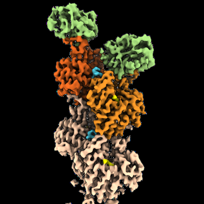
 Downloads & links
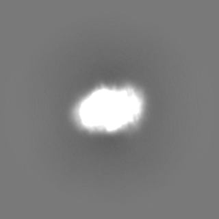
 emd_50516.png
 http://ftp.pdbj.org/pub/emdb/structures/EMD-50516
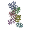


 Z (Sec.)
 Y (Row.)
 X (Col.)


 Sample components

 Processing
 Electron microscopy
 FIELD EMISSION GUN