 ムービー
ムービー コントローラー


+ データを開く

- 基本情報
基本情報
| 登録情報 | データベース: EMDB / ID: EMD-5040 | |||||||||
|---|---|---|---|---|---|---|---|---|---|---|
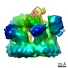
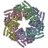
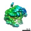
| タイトル | 3D structure of the ATP-bound yeast Vps4pE233Q dodecamer | |||||||||
 マップデータ マップデータ | 3D density map of ATP-bound yeast Vps4(delta)E233Q | |||||||||
 試料 試料 |
| |||||||||
 キーワード キーワード | protein transport / AAA ATPase / endosomal sorting / enveloped virus budding | |||||||||
| 手法 | 単粒子再構成法 / ネガティブ染色法 / 解像度: 23.0 Å | |||||||||
 データ登録者 データ登録者 | Landsberg MJ / Vajjhala PR / Rothnagel R / Munn AL / Hankamer B | |||||||||
 引用 引用 | ジャーナル: Structure / 年: 2009 タイトル: Three-dimensional structure of AAA ATPase Vps4: advancing structural insights into the mechanisms of endosomal sorting and enveloped virus budding. 著者: Michael John Landsberg / Parimala Rao Vajjhala / Rosalba Rothnagel / Alan Leslie Munn / Ben Hankamer /  要旨: Vps4 is a AAA ATPase that mediates endosomal membrane protein sorting. It is also a host factor hijacked by a diverse set of clinically important viruses, including HIV and Ebola, to facilitate viral ...Vps4 is a AAA ATPase that mediates endosomal membrane protein sorting. It is also a host factor hijacked by a diverse set of clinically important viruses, including HIV and Ebola, to facilitate viral budding. Here we present the three-dimensional structure of the hydrolysis-defective Vps4p(E233Q) mutant. Single-particle analysis, multiangle laser light scattering, and the docking of independently determined atomic models of Vps4 monomers reveal a complex with C6 point symmetry, distinguishing between a range of previously suggested oligomeric states (8-14 subunits). The 3D reconstruction also reveals a tail-to-tail subunit organization between the two rings of the complex and identifies the location of domains critical to complex assembly and interaction with partner proteins. Our refined Vps4 structure is better supported by independent lines of evidence than those previously proposed, and provides insights into the mechanism of endosomal membrane protein sorting and viral envelope budding. | |||||||||
| 履歴 |
|
- 構造の表示
構造の表示
| ムービー |
 ムービービューア ムービービューア |
|---|---|
| 構造ビューア | EMマップ: SurfViewMolmilJmol/JSmol |
| 添付画像 |

- ダウンロードとリンク
-EMDBアーカイブ
| マップデータ | emd_5040.map.gz | 2.4 MB | EMDBマップデータ形式 | |
|---|---|---|---|---|
| ヘッダ (付随情報) | emd-5040-v30.xmlemd-5040.xml | 9.7 KB 9.7 KB | 表示 表示 | EMDBヘッダ |
| 画像 |  emd_5040_1.png emd_5040_1.png | 154.3 KB | ||
| アーカイブディレクトリ |  http://ftp.pdbj.org/pub/emdb/structures/EMD-5040ftp://ftp.pdbj.org/pub/emdb/structures/EMD-5040 http://ftp.pdbj.org/pub/emdb/structures/EMD-5040ftp://ftp.pdbj.org/pub/emdb/structures/EMD-5040 | HTTPS FTP |
-関連構造データ


-リンク
| EMDBのページ | EMDB (EBI/PDBe) / EMDataResource |
|---|
-マップ
| ファイル | ダウンロード / ファイル: emd_5040.map.gz / 形式: CCP4 / 大きさ: 3.3 MB / タイプ: IMAGE STORED AS FLOATING POINT NUMBER (4 BYTES) | ||||||||||||||||||||||||||||||||||||||||||||||||||||||||||||||||||||
|---|---|---|---|---|---|---|---|---|---|---|---|---|---|---|---|---|---|---|---|---|---|---|---|---|---|---|---|---|---|---|---|---|---|---|---|---|---|---|---|---|---|---|---|---|---|---|---|---|---|---|---|---|---|---|---|---|---|---|---|---|---|---|---|---|---|---|---|---|---|
| 注釈 | 3D density map of ATP-bound yeast Vps4(delta)E233Q | ||||||||||||||||||||||||||||||||||||||||||||||||||||||||||||||||||||
| 投影像・断面図 | 画像のコントロール
画像は Spider により作成 | ||||||||||||||||||||||||||||||||||||||||||||||||||||||||||||||||||||
| ボクセルのサイズ | X=Y=Z: 4.087 Å | ||||||||||||||||||||||||||||||||||||||||||||||||||||||||||||||||||||
| 密度 |
| ||||||||||||||||||||||||||||||||||||||||||||||||||||||||||||||||||||
| 対称性 | 空間群: 1 | ||||||||||||||||||||||||||||||||||||||||||||||||||||||||||||||||||||
| 詳細 | EMDB XML:
CCP4マップ ヘッダ情報:
| ||||||||||||||||||||||||||||||||||||||||||||||||||||||||||||||||||||
 Z (Sec.)
 Y (Row.)
 X (Col.)


-添付データ
- 試料の構成要素
-全体 : 6xHis tagged yeast Vps4pE233Q
| 全体 | 名称: 6xHis tagged yeast Vps4pE233Q |
|---|---|
| 要素 |
|
-超分子 #1000: 6xHis tagged yeast Vps4pE233Q
| 超分子 | 名称: 6xHis tagged yeast Vps4pE233Q / タイプ: sample / ID: 1000 / 集合状態: The complex is a dodecamer / Number unique components: 1 |
|---|---|
| 分子量 | 実験値: 560 KDa / 理論値: 590 KDa / 手法: Multi-angle laser light scattering |
-分子 #1: Vacuolar protein sorting 4
| 分子 | 名称: Vacuolar protein sorting 4 / タイプ: protein_or_peptide / ID: 1 / Name.synonym: Vps4p(E233Q) / コピー数: 12 / 集合状態: Dodecamer / 組換発現: Yes |
|---|---|
| 由来(天然) | 株: Saccharomyces cerevisiae / 組織: cytosol / 細胞中の位置: Endosome membrame |
| 分子量 | 実験値: 560 KDa / 理論値: 590 KDa |
| 組換発現 | 生物種:  |
-実験情報
-構造解析
| 手法 | ネガティブ染色法 |
|---|---|
 解析 解析 | 単粒子再構成法 |
| 試料の集合状態 | particle |
-試料調製
| 濃度 | 0.1 mg/mL |
|---|---|
| 緩衝液 | pH: 7.4 詳細: 0.1M potassium acetate, 5mM magnesium acetate, 20mM Hepes, 1mM ATP |
| 染色 | タイプ: NEGATIVE 詳細: Grids with adsorbed protein washed with an aqueous solution of 2% (w/v) uranyl acetate for 30 seconds. |
| グリッド | 詳細: 400 mesh copper grid |
| 凍結 | 凍結剤: NONE / 装置: OTHER |
- 電子顕微鏡法
| 顕微鏡 | FEI TECNAI F30 |
|---|---|
| アライメント法 | Legacy - 非点収差: objective lens astigmatism was corrected at 93,000 times magnification |
| 撮影 | カテゴリ: CCD フィルム・検出器のモデル: GATAN ULTRASCAN 4000 (4k x 4k) 実像数: 170 / 平均電子線量: 60 e/Å2 / ビット/ピクセル: 8 |
| 電子線 | 加速電圧: 300 kV / 電子線源:  FIELD EMISSION GUN FIELD EMISSION GUN |
| 電子光学系 | 照射モード: FLOOD BEAM / 撮影モード: BRIGHT FIELD / 最大 デフォーカス(公称値): 1.1 µm / 最小 デフォーカス(公称値): 0.6 µm / 倍率(公称値): 59000 |
| 試料ステージ | 試料ホルダー: single tilt / 試料ホルダーモデル: OTHER |
| 実験機器 |  モデル: Tecnai F30 / 画像提供: FEI Company |
-画像解析
| 詳細 | Particles were selected semi-interactively using SwarmPS |
|---|---|
| 最終 再構成 | アルゴリズム: OTHER / 解像度のタイプ: BY AUTHOR / 解像度: 23.0 Å / 解像度の算出法: OTHER / ソフトウェア - 名称: IMAGIC, XMIPP, EMAN / 詳細: FSC at 0.5 cut-off estimates resolution at 18.3 / 使用した粒子像数: 8590 |
| 最終 2次元分類 | クラス数: 340 |
