 Movie
Movie Controller


+ Open data

- Basic information
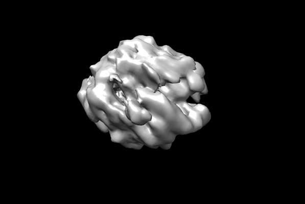
| Entry | Database: EMDB / ID: EMD-8885 | |||||||||
|---|---|---|---|---|---|---|---|---|---|---|
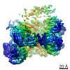
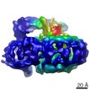
| Title | RNA Pol II complex arrested by a CPD lesion | |||||||||
 Map data Map data | RNA Pol II elongation complex with a CPD lesion containing scaffold | |||||||||
 Sample Sample |
| |||||||||
| Biological species |  | |||||||||
| Method | single particle reconstruction / cryo EM / Resolution: 10.0 Å | |||||||||
 Authors Authors | Lahiri I / Leschziner AE | |||||||||
| Funding support |  United States, 1 items United States, 1 items
| |||||||||
 Citation Citation | Journal: Nature / Year: 2017 Title: Structural basis for the initiation of eukaryotic transcription-coupled DNA repair. Authors: Jun Xu / Indrajit Lahiri / Wei Wang / Adam Wier / Michael A Cianfrocco / Jenny Chong / Alissa A Hare / Peter B Dervan / Frank DiMaio / Andres E Leschziner / Dong Wang / Abstract: Eukaryotic transcription-coupled repair (TCR) is an important and well-conserved sub-pathway of nucleotide excision repair that preferentially removes DNA lesions from the template strand that block ...Eukaryotic transcription-coupled repair (TCR) is an important and well-conserved sub-pathway of nucleotide excision repair that preferentially removes DNA lesions from the template strand that block translocation of RNA polymerase II (Pol II). Cockayne syndrome group B (CSB, also known as ERCC6) protein in humans (or its yeast orthologues, Rad26 in Saccharomyces cerevisiae and Rhp26 in Schizosaccharomyces pombe) is among the first proteins to be recruited to the lesion-arrested Pol II during the initiation of eukaryotic TCR. Mutations in CSB are associated with the autosomal-recessive neurological disorder Cockayne syndrome, which is characterized by progeriod features, growth failure and photosensitivity. The molecular mechanism of eukaryotic TCR initiation remains unclear, with several long-standing unanswered questions. How cells distinguish DNA lesion-arrested Pol II from other forms of arrested Pol II, the role of CSB in TCR initiation, and how CSB interacts with the arrested Pol II complex are all unknown. The lack of structures of CSB or the Pol II-CSB complex has hindered our ability to address these questions. Here we report the structure of the S. cerevisiae Pol II-Rad26 complex solved by cryo-electron microscopy. The structure reveals that Rad26 binds to the DNA upstream of Pol II, where it markedly alters its path. Our structural and functional data suggest that the conserved Swi2/Snf2-family core ATPase domain promotes the forward movement of Pol II, and elucidate key roles for Rad26 in both TCR and transcription elongation. | |||||||||
| History |
|
- Structure visualization
| Movie |
 Movie viewer Movie viewer |
|---|---|
| Structure viewer | EM map: SurfViewMolmilJmol/JSmol |
| Supplemental images |
- Downloads & links
-EMDB archive
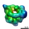
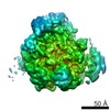
| Map data | emd_8885.map.gz | 4.1 MB | EMDB map data format | |
|---|---|---|---|---|
| Header (meta data) | emd-8885-v30.xmlemd-8885.xml | 9 KB 9 KB | Display Display | EMDB header |
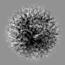
| Images |  emd_8885.png emd_8885.png | 37.5 KB | ||
| Archive directory |  http://ftp.pdbj.org/pub/emdb/structures/EMD-8885ftp://ftp.pdbj.org/pub/emdb/structures/EMD-8885 http://ftp.pdbj.org/pub/emdb/structures/EMD-8885ftp://ftp.pdbj.org/pub/emdb/structures/EMD-8885 | HTTPS FTP |
-Related structure data
| Related structure data |  7038C  8735C 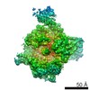 8736C 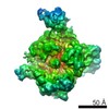 8737C  5vvrC 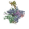 5vvsC C: citing same article ( |
|---|---|
| Similar structure data |


-Links
| EMDB pages | EMDB (EBI/PDBe) / EMDataResource |
|---|
-Map
| File | Download / File: emd_8885.map.gz / Format: CCP4 / Size: 8 MB / Type: IMAGE STORED AS FLOATING POINT NUMBER (4 BYTES) | ||||||||||||||||||||||||||||||||||||||||||||||||||||||||||||||||||||
|---|---|---|---|---|---|---|---|---|---|---|---|---|---|---|---|---|---|---|---|---|---|---|---|---|---|---|---|---|---|---|---|---|---|---|---|---|---|---|---|---|---|---|---|---|---|---|---|---|---|---|---|---|---|---|---|---|---|---|---|---|---|---|---|---|---|---|---|---|---|
| Annotation | RNA Pol II elongation complex with a CPD lesion containing scaffold | ||||||||||||||||||||||||||||||||||||||||||||||||||||||||||||||||||||
| Projections & slices | Image control
Images are generated by Spider. | ||||||||||||||||||||||||||||||||||||||||||||||||||||||||||||||||||||
| Voxel size | X=Y=Z: 3.6 Å | ||||||||||||||||||||||||||||||||||||||||||||||||||||||||||||||||||||
| Density |
| ||||||||||||||||||||||||||||||||||||||||||||||||||||||||||||||||||||
| Symmetry | Space group: 1 | ||||||||||||||||||||||||||||||||||||||||||||||||||||||||||||||||||||
| Details | EMDB XML:
CCP4 map header:
| ||||||||||||||||||||||||||||||||||||||||||||||||||||||||||||||||||||
 Z (Sec.)
 Y (Row.)
 X (Col.)


-Supplemental data
- Sample components
-Entire : Binary complex of RNA Pol II and transcription scaffold
| Entire | Name: Binary complex of RNA Pol II and transcription scaffold |
|---|---|
| Components |
|
-Supramolecule #1: Binary complex of RNA Pol II and transcription scaffold
| Supramolecule | Name: Binary complex of RNA Pol II and transcription scaffold type: complex / ID: 1 / Parent: 0 |
|---|---|
| Source (natural) | Organism: Strain: ATCC 204508 / S288c |
-Experimental details
-Structure determination
| Method | cryo EM |
|---|---|
 Processing Processing | single particle reconstruction |
| Aggregation state | particle |
-Sample preparation
| Buffer | pH: 7.5 |
|---|---|
| Vitrification | Cryogen name: ETHANE / Chamber humidity: 90 % / Chamber temperature: 277.15 K / Instrument: FEI VITROBOT MARK IV |
- Electron microscopy
| Microscope | FEI TALOS ARCTICA |
|---|---|
| Image recording | Film or detector model: GATAN K2 SUMMIT (4k x 4k) / Detector mode: SUPER-RESOLUTION / Average electron dose: 7.7 e/Å2 |
| Electron beam | Acceleration voltage: 200 kV / Electron source:  FIELD EMISSION GUN FIELD EMISSION GUN |
| Electron optics | Illumination mode: FLOOD BEAM / Imaging mode: BRIGHT FIELD |
| Experimental equipment |  Model: Talos Arctica / Image courtesy: FEI Company |
-Image processing
| Startup model | Type of model: PDB ENTRY PDB model - PDB ID: |
|---|---|
| Final reconstruction | Applied symmetry - Point group: C1 (asymmetric) / Resolution.type: BY AUTHOR / Resolution: 10.0 Å / Resolution method: FSC 0.5 CUT-OFF / Software - Name: cryoSPARC Details: The particles contributing to the structure adopted a strong preferred orientation. Number images used: 51119 |
| Initial angle assignment | Type: PROJECTION MATCHING |
| Final angle assignment | Type: PROJECTION MATCHING |
