 Movie
Movie Controller


+ Open data

- Basic information
| Entry | Database: EMDB / ID: EMD-5037 | |||||||||
|---|---|---|---|---|---|---|---|---|---|---|
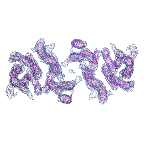
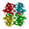
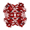
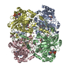
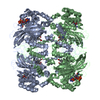
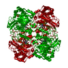
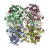
| Title | 3D EM map of E.coli NhaA | |||||||||
 Map data Map data | NhaA | |||||||||
 Sample Sample |
| |||||||||
 Keywords Keywords | Sodium proton antiporter | |||||||||
| Function / homology |  Function and homology information Function and homology informationresponse to alkaline pH / sodium:proton antiporter activity / cardiolipin binding / response to salt stress / regulation of intracellular pH / plasma membrane Similarity search - Function | |||||||||
| Biological species |  | |||||||||
| Method | electron crystallography / cryo EM | |||||||||
 Authors Authors | Williams K / Kuehlbrandt W | |||||||||
 Citation Citation | Journal: Nature / Year: 2000 Title: Three-dimensional structure of the ion-coupled transport protein NhaA. Authors: K A Williams /  Abstract: Ion-coupled membrane-transport proteins, or secondary transporters, comprise a diverse and abundant group of membrane proteins that are found in all organisms. These proteins facilitate solute ...Ion-coupled membrane-transport proteins, or secondary transporters, comprise a diverse and abundant group of membrane proteins that are found in all organisms. These proteins facilitate solute accumulation and toxin removal against concentration gradients using energy supplied by ion gradients across membranes. NhaA is a Na+/H+ antiporter of relative molecular mass 42,000, which is found in the inner membrane of Escherichia coli, and which has been cloned and characterized. NhaA uses the H+ electrochemical gradient to expel Na+ from the cytoplasm, and functions primarily in the adaptation to high salinity at alkaline pH. Most secondary transporters, including NhaA, are predicted to have 12 transmembrane helices. Here we report the structure of NhaA, at 7 A resolution in the membrane plane and at 14 A vertical resolution, determined from two-dimensional crystals using electron cryo-microscopy. The three-dimensional map of NhaA reveals 12 tilted, bilayer-spanning helices. A roughly linear arrangement of six helices is adjacent to a compact bundle of six helices, with the density for one helix in the bundle not continuous through the membrane. The molecular organization of NhaA represents a new membrane-protein structural motif and offers the first insights into the architecture of an ion-coupled transport protein. | |||||||||
| History |
|
- Structure visualization
| Movie |
Movie viewer |
|---|---|
| Structure viewer | EM map: SurfViewMolmilJmol/JSmol |
| Supplemental images |
- Downloads & links
-EMDB archive
| Map data | emd_5037.map.gz | 187.8 KB | EMDB map data format | |
|---|---|---|---|---|
| Header (meta data) | emd-5037-v30.xmlemd-5037.xml | 8.1 KB 8.1 KB | Display Display | EMDB header |
| Images |  emd_5037_1.png emd_5037_1.png | 173.5 KB | ||
| Archive directory |  http://ftp.pdbj.org/pub/emdb/structures/EMD-5037ftp://ftp.pdbj.org/pub/emdb/structures/EMD-5037 http://ftp.pdbj.org/pub/emdb/structures/EMD-5037ftp://ftp.pdbj.org/pub/emdb/structures/EMD-5037 | HTTPS FTP |
-Validation report
| Summary document | emd_5037_validation.pdf.gz | 314.6 KB | Display | EMDB validaton report |
|---|---|---|---|---|
| Full document | emd_5037_full_validation.pdf.gz | 314.2 KB | Display | |
| Data in XML | emd_5037_validation.xml.gz | 4.6 KB | Display | |
| Arichive directory | https://ftp.pdbj.org/pub/emdb/validation_reports/EMD-5037ftp://ftp.pdbj.org/pub/emdb/validation_reports/EMD-5037 | HTTPS FTP |
-Related structure data
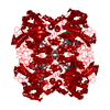
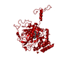
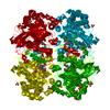
| Related structure data |  3fi1M M: atomic model generated by this map |
|---|---|
| Similar structure data |

-Links
| EMDB pages | EMDB (EBI/PDBe) / EMDataResource |
|---|---|
| Related items in Molecule of the Month |

-Map
| File | Download / File: emd_5037.map.gz / Format: CCP4 / Size: 335 KB / Type: IMAGE STORED AS FLOATING POINT NUMBER (4 BYTES) | ||||||||||||||||||||||||||||||||||||||||||||||||||||||||||||||||||||
|---|---|---|---|---|---|---|---|---|---|---|---|---|---|---|---|---|---|---|---|---|---|---|---|---|---|---|---|---|---|---|---|---|---|---|---|---|---|---|---|---|---|---|---|---|---|---|---|---|---|---|---|---|---|---|---|---|---|---|---|---|---|---|---|---|---|---|---|---|---|
| Annotation | NhaA | ||||||||||||||||||||||||||||||||||||||||||||||||||||||||||||||||||||
| Projections & slices | Image control
Images are generated by Spider. generated in cubic-lattice coordinate | ||||||||||||||||||||||||||||||||||||||||||||||||||||||||||||||||||||
| Voxel size | X: 1.97917 Å / Y: 2.06136 Å / Z: 2.08333 Å | ||||||||||||||||||||||||||||||||||||||||||||||||||||||||||||||||||||
| Density |
| ||||||||||||||||||||||||||||||||||||||||||||||||||||||||||||||||||||
| Symmetry | Space group: 18 | ||||||||||||||||||||||||||||||||||||||||||||||||||||||||||||||||||||
| Details | EMDB XML:
CCP4 map header:
| ||||||||||||||||||||||||||||||||||||||||||||||||||||||||||||||||||||
 Y (Sec.)
 X (Row.)
 Z (Col.)


-Supplemental data
- Sample components
-Entire : 3D EM map of NhaA 2D crystals
| Entire | Name: 3D EM map of NhaA 2D crystals |
|---|---|
| Components |
|
-Supramolecule #1000: 3D EM map of NhaA 2D crystals
| Supramolecule | Name: 3D EM map of NhaA 2D crystals / type: sample / ID: 1000 / Details: 2D crystals / Oligomeric state: Dimer / Number unique components: 2 |
|---|
-Supramolecule #1: NhaA
| Supramolecule | Name: NhaA / type: organelle_or_cellular_component / ID: 1 / Name.synonym: NhaA / Oligomeric state: dimer / Recombinant expression: Yes |
|---|---|
| Source (natural) | Organism: |
| Recombinant expression | Organism: |
-Experimental details
-Structure determination
| Method | cryo EM |
|---|---|
 Processing Processing | electron crystallography |
| Aggregation state | 2D array |
-Sample preparation
| Concentration | 0.5 mg/mL |
|---|---|
| Vitrification | Cryogen name: NITROGEN / Instrument: OTHER |
- Electron microscopy
| Microscope | JEOL 3000SFF |
|---|---|
| Temperature | Min: 4 K |
| Image recording | Category: FILM / Film or detector model: KODAK SO-163 FILM / Digitization - Scanner: ZEISS SCAI / Digitization - Sampling interval: 7 µm |
| Tilt angle min | 0 |
| Electron beam | Acceleration voltage: 300 kV / Electron source:  FIELD EMISSION GUN FIELD EMISSION GUN |
| Electron optics | Illumination mode: OTHER / Imaging mode: DIFFRACTION |
| Sample stage | Specimen holder: Holder / Specimen holder model: OTHER / Tilt angle max: 45 / Tilt series - Axis1 - Min angle: 0 ° / Tilt series - Axis1 - Max angle: 45 ° |
-Image processing
| Details | ab is plane as defined by unit cell parameters (s.g P 21 21 2) |
|---|---|
| Final reconstruction | Software - Name: CCP4 |
| Crystal parameters | Unit cell - A: 47.50 Å / Unit cell - B: 181.40 Å / Unit cell - C: 200.00 Å / Unit cell - γ: 90.00 ° / Unit cell - α: 90.00 ° / Unit cell - β: 90.00 ° / Plane group: P 2 21 21 |
