: / CENP-A recruiting complex / chromocenter / cell communication / CENP-A containing chromatin assembly / protein localization to chromosome, centromeric region / chromosome, centromeric region / Cajal body / Cyclin A/B1/B2 associated events during G2/M transition / Deposition of new CENPA-containing nucleosomes at the centromere ...: / CENP-A recruiting complex / chromocenter / cell communication / CENP-A containing chromatin assembly / protein localization to chromosome, centromeric region / chromosome, centromeric region / Cajal body / Cyclin A/B1/B2 associated events during G2/M transition / Deposition of new CENPA-containing nucleosomes at the centromere / chromosome segregation / protein-macromolecule adaptor activity / nuclear speck / cell division / chromatin / DNA binding / nucleoplasm / metal ion binding / identical protein binding / nucleus / cytosol Similarity search - Function
Mis18 domain / Mis18 domain profile. / SANT associated / KNL2-like / SANTA (SANT Associated) / Yippee/Mis18/Cereblon / Yippee zinc-binding/DNA-binding /Mis18, centromere assembly / SANT domain profile. / SANT domain / SANT SWI3, ADA2, N-CoR and TFIIIB'' DNA-binding domains ...Mis18 domain / Mis18 domain profile. / SANT associated / KNL2-like / SANTA (SANT Associated) / Yippee/Mis18/Cereblon / Yippee zinc-binding/DNA-binding /Mis18, centromere assembly / SANT domain profile. / SANT domain / SANT SWI3, ADA2, N-CoR and TFIIIB'' DNA-binding domains / SANT/Myb domain / Homeobox-like domain superfamily Similarity search - Domain/homology
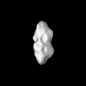
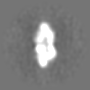
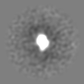
Journal: EMBO Rep / Year: 2024 Title: Structural basis for Mis18 complex assembly and its implications for centromere maintenance. Authors: Reshma Thamkachy / Bethan Medina-Pritchard / Sang Ho Park / Carla G Chiodi / Juan Zou / Maria de la Torre-Barranco / Kazuma Shimanaka / Maria Alba Abad / Cristina Gallego Páramo / Regina ...Authors: Reshma Thamkachy / Bethan Medina-Pritchard / Sang Ho Park / Carla G Chiodi / Juan Zou / Maria de la Torre-Barranco / Kazuma Shimanaka / Maria Alba Abad / Cristina Gallego Páramo / Regina Feederle / Emilija Ruksenaite / Patrick Heun / Owen R Davies / Juri Rappsilber / Dina Schneidman-Duhovny / Uhn-Soo Cho / A Arockia Jeyaprakash / Abstract: The centromere, defined by the enrichment of CENP-A (a Histone H3 variant) containing nucleosomes, is a specialised chromosomal locus that acts as a microtubule attachment site. To preserve ...The centromere, defined by the enrichment of CENP-A (a Histone H3 variant) containing nucleosomes, is a specialised chromosomal locus that acts as a microtubule attachment site. To preserve centromere identity, CENP-A levels must be maintained through active CENP-A loading during the cell cycle. A central player mediating this process is the Mis18 complex (Mis18α, Mis18β and Mis18BP1), which recruits the CENP-A-specific chaperone HJURP to centromeres for CENP-A deposition. Here, using a multi-pronged approach, we characterise the structure of the Mis18 complex and show that multiple hetero- and homo-oligomeric interfaces facilitate the hetero-octameric Mis18 complex assembly composed of 4 Mis18α, 2 Mis18β and 2 Mis18BP1. Evaluation of structure-guided/separation-of-function mutants reveals structural determinants essential for cell cycle controlled Mis18 complex assembly and centromere maintenance. Our results provide new mechanistic insights on centromere maintenance, highlighting that while Mis18α can associate with centromeres and deposit CENP-A independently of Mis18β, the latter is indispensable for the optimal level of CENP-A loading required for preserving the centromere identity.
In the structure databanks used in Yorodumi, some data are registered as the other names, "COVID-19 virus" and "2019-nCoV". Here are the details of the virus and the list of structure data.
Jan 31, 2019. EMDB accession codes are about to change! (news from PDBe EMDB page)
EMDB accession codes are about to change! (news from PDBe EMDB page)
The allocation of 4 digits for EMDB accession codes will soon come to an end. Whilst these codes will remain in use, new EMDB accession codes will include an additional digit and will expand incrementally as the available range of codes is exhausted. The current 4-digit format prefixed with “EMD-” (i.e. EMD-XXXX) will advance to a 5-digit format (i.e. EMD-XXXXX), and so on. It is currently estimated that the 4-digit codes will be depleted around Spring 2019, at which point the 5-digit format will come into force.
The EM Navigator/Yorodumi systems omit the EMD- prefix.
Related info.:Q: What is EMD? / ID/Accession-code notation in Yorodumi/EM Navigator
Yorodumi is a browser for structure data from EMDB, PDB, SASBDB, etc.
This page is also the successor to EM Navigator detail page, and also detail information page/front-end page for Omokage search.
The word "yorodu" (or yorozu) is an old Japanese word meaning "ten thousand". "mi" (miru) is to see.
Related info.:EMDB / PDB / SASBDB / Comparison of 3 databanks / Yorodumi Search / Aug 31, 2016. New EM Navigator & Yorodumi / Yorodumi Papers / Jmol/JSmol / Function and homology information / Changes in new EM Navigator and Yorodumi
 Movie
Movie Controller


 Open data
 Basic information
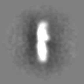
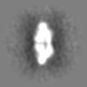
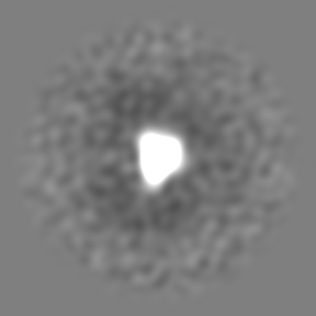
 Map data
 Sample
 Keywords
 Function and homology information
 Homo sapiens (human)
 Authors
 United Kingdom, European Union, 3 items
  Citation


 Structure visualization
 Downloads & links
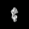
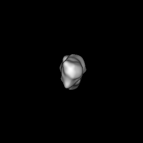
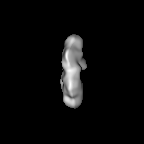
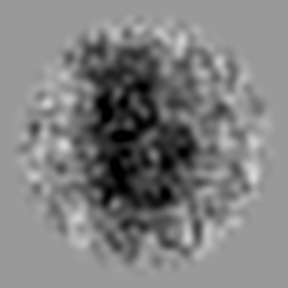
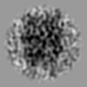
 emd_50219.png
 http://ftp.pdbj.org/pub/emdb/structures/EMD-50219


 Z (Sec.)
 Y (Row.)
 X (Col.)


 Sample components
 Processing
 Electron microscopy
 FIELD EMISSION GUN