ムービー
ムービー コントローラー


+ データを開く

- 基本情報
基本情報
| 登録情報 | データベース: EMDB / ID: EMD-5016 | |||||||||
|---|---|---|---|---|---|---|---|---|---|---|
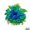
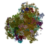
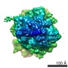
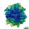
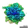
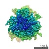
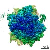
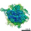
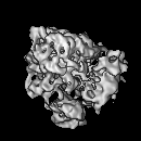
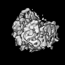
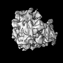
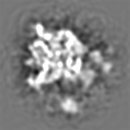
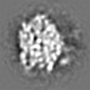
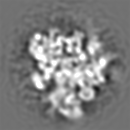
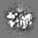
| タイトル | 15.3A eEF2-80S Ribosome Transition State Complex by Cryo-electron Microscopy | |||||||||
 マップデータ マップデータ | 15.3A eEF2.80S cryo-EM map | |||||||||
 試料 試料 |
| |||||||||
 キーワード キーワード | AlF4- / GDP / GTPase / Ribosome / Translocation | |||||||||
| 生物種 |   Thermomyces lanuginosus (菌類) Thermomyces lanuginosus (菌類) | |||||||||
| 手法 | 単粒子再構成法 / クライオ電子顕微鏡法 / 解像度: 15.3 Å | |||||||||
 データ登録者 データ登録者 | Sengupta J / Nilsson J / Gursky R / Kjeldgaard M / Nissen P / Frank J | |||||||||
 引用 引用 | ジャーナル: J Mol Biol / 年: 2008 タイトル: Visualization of the eEF2-80S ribosome transition-state complex by cryo-electron microscopy. 著者: Jayati Sengupta / Jakob Nilsson / Richard Gursky / Morten Kjeldgaard / Poul Nissen / Joachim Frank /  要旨: In an attempt to understand ribosome-induced GTP hydrolysis on eEF2, we determined a 12.6-A cryo-electron microscopy reconstruction of the eEF2-bound 80S ribosome in the presence of aluminum ...In an attempt to understand ribosome-induced GTP hydrolysis on eEF2, we determined a 12.6-A cryo-electron microscopy reconstruction of the eEF2-bound 80S ribosome in the presence of aluminum tetrafluoride and GDP, with aluminum tetrafluoride mimicking the gamma-phosphate during hydrolysis. This is the first visualization of a structure representing a transition-state complex on the ribosome. Tight interactions are observed between the factor's G domain and the large ribosomal subunit, as well as between domain IV and an intersubunit bridge. In contrast, some of the domains of eEF2 implicated in small subunit binding display a large degree of flexibility. Furthermore, we find support for a transition-state model conformation of the switch I region in this complex where the reoriented switch I region interacts with a conserved rRNA region of the 40S subunit formed by loops of the 18S RNA helices 8 and 14. This complex is structurally distinct from the eEF2-bound 80S ribosome complexes previously reported, and analysis of this map sheds light on the GTPase-coupled translocation mechanism. | |||||||||
| 履歴 |
|
- 構造の表示
構造の表示
| ムービー |
 ムービービューア ムービービューア |
|---|---|
| 構造ビューア | EMマップ: SurfViewMolmilJmol/JSmol |
| 添付画像 |

- ダウンロードとリンク
-EMDBアーカイブ
| マップデータ | emd_5016.map.gz | 7.9 MB | EMDBマップデータ形式 | |
|---|---|---|---|---|
| ヘッダ (付随情報) | emd-5016-v30.xmlemd-5016.xml | 9.3 KB 9.3 KB | 表示 表示 | EMDBヘッダ |
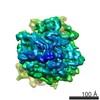
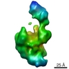
| 画像 | 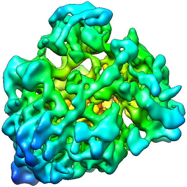 emd_5016_1.png emd_5016_1.png | 315.9 KB | ||
| アーカイブディレクトリ |  http://ftp.pdbj.org/pub/emdb/structures/EMD-5016ftp://ftp.pdbj.org/pub/emdb/structures/EMD-5016 http://ftp.pdbj.org/pub/emdb/structures/EMD-5016ftp://ftp.pdbj.org/pub/emdb/structures/EMD-5016 | HTTPS FTP |
-検証レポート
| 文書・要旨 | emd_5016_validation.pdf.gz | 78.3 KB | 表示 | EMDB検証レポート |
|---|---|---|---|---|
| 文書・詳細版 | emd_5016_full_validation.pdf.gz | 77.4 KB | 表示 | |
| XML形式データ | emd_5016_validation.xml.gz | 494 B | 表示 | |
| アーカイブディレクトリ | https://ftp.pdbj.org/pub/emdb/validation_reports/EMD-5016ftp://ftp.pdbj.org/pub/emdb/validation_reports/EMD-5016 | HTTPS FTP |
-関連構造データ
-リンク
| EMDBのページ | EMDB (EBI/PDBe) / EMDataResource |
|---|---|
| 「今月の分子」の関連する項目 |

-マップ
| ファイル | ダウンロード / ファイル: emd_5016.map.gz / 形式: CCP4 / 大きさ: 8.2 MB / タイプ: IMAGE STORED AS FLOATING POINT NUMBER (4 BYTES) | ||||||||||||||||||||||||||||||||||||||||||||||||||||||||||||||||||||
|---|---|---|---|---|---|---|---|---|---|---|---|---|---|---|---|---|---|---|---|---|---|---|---|---|---|---|---|---|---|---|---|---|---|---|---|---|---|---|---|---|---|---|---|---|---|---|---|---|---|---|---|---|---|---|---|---|---|---|---|---|---|---|---|---|---|---|---|---|---|
| 注釈 | 15.3A eEF2.80S cryo-EM map | ||||||||||||||||||||||||||||||||||||||||||||||||||||||||||||||||||||
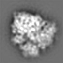
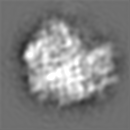
| 投影像・断面図 | 画像のコントロール
画像は Spider により作成 | ||||||||||||||||||||||||||||||||||||||||||||||||||||||||||||||||||||
| ボクセルのサイズ | X=Y=Z: 2.82 Å | ||||||||||||||||||||||||||||||||||||||||||||||||||||||||||||||||||||
| 密度 |
| ||||||||||||||||||||||||||||||||||||||||||||||||||||||||||||||||||||
| 対称性 | 空間群: 1 | ||||||||||||||||||||||||||||||||||||||||||||||||||||||||||||||||||||
| 詳細 | EMDB XML:
CCP4マップ ヘッダ情報:
| ||||||||||||||||||||||||||||||||||||||||||||||||||||||||||||||||||||
 Z (Sec.)
 Y (Row.)
 X (Col.)


-添付データ
- 試料の構成要素
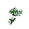
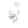
-全体 : 80S ribosome in complex with eEF2
| 全体 | 名称: 80S ribosome in complex with eEF2 |
|---|---|
| 要素 |
|
-超分子 #1000: 80S ribosome in complex with eEF2
| 超分子 | 名称: 80S ribosome in complex with eEF2 / タイプ: sample / ID: 1000 / Number unique components: 4 |
|---|
-超分子 #1: 80S ribosome
| 超分子 | 名称: 80S ribosome / タイプ: complex / ID: 1 / 組換発現: No / データベース: NCBI / Ribosome-details: ribosome-eukaryote: ALL |
|---|---|
| 由来(天然) | 生物種: Thermomyces lanuginosus (菌類) |
-実験情報
-構造解析
| 手法 | クライオ電子顕微鏡法 |
|---|---|
 解析 解析 | 単粒子再構成法 |
| 試料の集合状態 | particle |
-試料調製
| 緩衝液 | pH: 7.2 / 詳細: 20 mM Hepes-NH3, 100 mM KCl, 20 mM MgCl2 |
|---|---|
| グリッド | 詳細: Quantifoil grid |
| 凍結 | 凍結剤: ETHANE / チャンバー内湿度: 90 % / チャンバー内温度: 93 K / 装置: OTHER / 詳細: Vitrification instrument: Vitrobot |
- 電子顕微鏡法
| 顕微鏡 | FEI TECNAI F20 |
|---|---|
| 撮影 | デジタル化 - サンプリング間隔: 2.82 µm / 平均電子線量: 10 e/Å2 / ビット/ピクセル: 16 |
| 電子線 | 加速電圧: 200 kV / 電子線源:  FIELD EMISSION GUN FIELD EMISSION GUN |
| 電子光学系 | 照射モード: FLOOD BEAM / 撮影モード: BRIGHT FIELD / 最大 デフォーカス(公称値): 4.5 µm / 最小 デフォーカス(公称値): 1.5 µm / 倍率(公称値): 50000 |
| 試料ステージ | 試料ホルダー: Single tilt cyro-holder / 試料ホルダーモデル: GATAN LIQUID NITROGEN |
| 実験機器 |  モデル: Tecnai F20 / 画像提供: FEI Company |
-画像解析
| 詳細 | The image processing using SPIDER included a 3D projection alignment procedure with correction of the contrast transfer function and enhancement of the high-resolution Fourier amplitudes based on X-ray solution scattering data. |
|---|---|
| CTF補正 | 詳細: Segregation in defocus groups and correction in volumes |
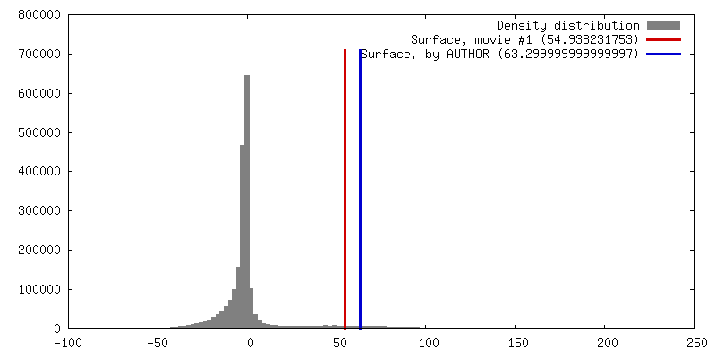
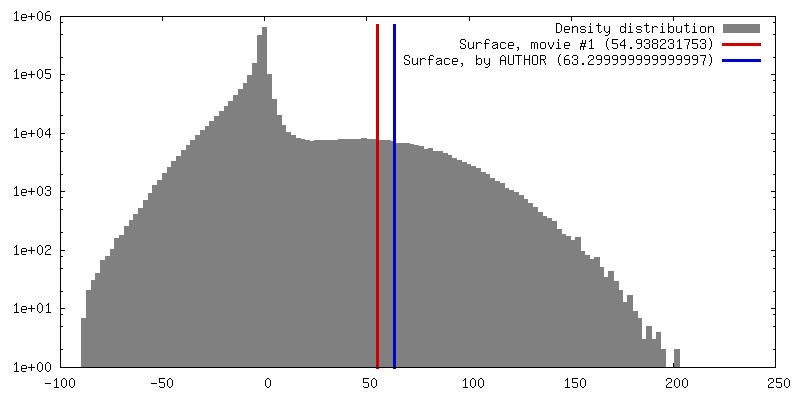
| 最終 再構成 | アルゴリズム: OTHER / 解像度のタイプ: BY AUTHOR / 解像度: 15.3 Å / 解像度の算出法: FSC 0.5 CUT-OFF / ソフトウェア - 名称: Spider 詳細: Supervised classification was used. Application of a more rigorous classification using smaller subset of particles (18,221) further improves the homogeneity of the complex, where the resolution changed to 15.3A. 使用した粒子像数: 18221 |
-原子モデル構築 1
| 初期モデル | PDB ID: |
|---|---|
| 詳細 | The domains were separately fitted by manual docking using program O |
| 精密化 | 空間: REAL / プロトコル: RIGID BODY FIT / 当てはまり具合の基準: correlation coefficient |