 Movie
Movie Controller

[English] 日本語
 Yorodumi
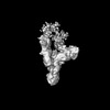
- EMDB-50105: Initial 3D Map of relaxosome complex with oriT DNA ds-67_+113(pol... -
+ Open data

- Basic information
| Entry |  | |||||||||
|---|---|---|---|---|---|---|---|---|---|---|
| Title | Initial 3D Map of relaxosome complex with oriT DNA ds-67_+113(poly-dT15-17_-3)deltaTraM | |||||||||
 Map data Map data | 3D refinement map Unsharpened | |||||||||
 Sample Sample |
| |||||||||
 Keywords Keywords | Relaxosome / Bacterial Conjugation / DNA processing / Relaxase / DNA binding proteins / DNA BINDING PROTEIN | |||||||||
| Biological species |  | |||||||||
| Method | single particle reconstruction / cryo EM / Resolution: 4.43 Å | |||||||||
 Authors Authors | Williams SM / Waksman G | |||||||||
| Funding support |  United Kingdom, 1 items United Kingdom, 1 items
| |||||||||
 Citation Citation | Journal: Nat Commun / Year: 2025 Title: Cryo-EM Structure of the relaxosome, a complex essential for bacterial mating and the spread of antibiotic resistance genes. Authors: Sunanda M Williams / Sandra Raffl / Sabine Kienesberger / Aravindan Ilangovan / Ellen L Zechner / Gabriel Waksman /  Abstract: Bacterial mating, or conjugation, was discovered nearly 80 years ago as a process transferring genes from one bacterial cell (the donor) to another (the recipient). It requires three key multiprotein ...Bacterial mating, or conjugation, was discovered nearly 80 years ago as a process transferring genes from one bacterial cell (the donor) to another (the recipient). It requires three key multiprotein complexes in the donor cell: a DNA-processing machinery called the relaxosome, a double-membrane spanning type 4 secretion system (T4SS), and an extracellular appendage termed pilus. While the near-atomic resolution structures of the T4SS and pilus are already known, that of the relaxosome has not been reported to date. Here, we describe the cryo-EM structure of the fully assembled relaxosome encoded by the paradigm F plasmid in two different states corresponding to distinct functional steps along the DNA processing reaction. By varying the structures of model DNAs we delineate conformational changes required to initiate conjugation. Mutational studies of the various protein-protein and protein-DNA interaction hubs suggest a complex sensitive to trigger signals, that could arise from cell-to-cell contacts with recipient cells. | |||||||||
| History |
|
- Structure visualization
| Supplemental images |
|---|

- Downloads & links
-EMDB archive
| Map data | emd_50105.map.gz | 140.3 MB |  EMDB map data format EMDB map data format | |
|---|---|---|---|---|
| Header (meta data) | emd-50105-v30.xmlemd-50105.xml | 17.5 KB 17.5 KB | Display Display | EMDB header |
| FSC (resolution estimation) | emd_50105_fsc.xml | 14 KB | Display | FSC data file |
| Images |  emd_50105.png emd_50105.png | 54.7 KB | ||
| Filedesc metadata | emd-50105.cif.gz | 4.6 KB | ||
| Others | emd_50105_additional_1.map.gzemd_50105_half_map_1.map.gzemd_50105_half_map_2.map.gz | 264.6 MB 262 MB 262 MB | ||
| Archive directory |  http://ftp.pdbj.org/pub/emdb/structures/EMD-50105ftp://ftp.pdbj.org/pub/emdb/structures/EMD-50105 http://ftp.pdbj.org/pub/emdb/structures/EMD-50105ftp://ftp.pdbj.org/pub/emdb/structures/EMD-50105 | HTTPS FTP |
-Validation report
| Summary document | emd_50105_validation.pdf.gz | 919.4 KB | Display | EMDB validaton report |
|---|---|---|---|---|
| Full document | emd_50105_full_validation.pdf.gz | 919.2 KB | Display | |
| Data in XML | emd_50105_validation.xml.gz | 23.2 KB | Display | |
| Data in CIF | emd_50105_validation.cif.gz | 30.1 KB | Display | |
| Arichive directory | https://ftp.pdbj.org/pub/emdb/validation_reports/EMD-50105ftp://ftp.pdbj.org/pub/emdb/validation_reports/EMD-50105 | HTTPS FTP |
-Related structure data


-Links
| EMDB pages | EMDB (EBI/PDBe) / EMDataResource |
|---|
-Map
| File | Download / File: emd_50105.map.gz / Format: CCP4 / Size: 282.6 MB / Type: IMAGE STORED AS FLOATING POINT NUMBER (4 BYTES) | ||||||||||||||||||||||||||||||||||||
|---|---|---|---|---|---|---|---|---|---|---|---|---|---|---|---|---|---|---|---|---|---|---|---|---|---|---|---|---|---|---|---|---|---|---|---|---|---|
| Annotation | 3D refinement map Unsharpened | ||||||||||||||||||||||||||||||||||||
| Projections & slices | Image control
Images are generated by Spider. | ||||||||||||||||||||||||||||||||||||
| Voxel size | X=Y=Z: 0.828 Å | ||||||||||||||||||||||||||||||||||||
| Density |
| ||||||||||||||||||||||||||||||||||||
| Symmetry | Space group: 1 | ||||||||||||||||||||||||||||||||||||
| Details | EMDB XML:
|
 Z (Sec.)
 Y (Row.)
 X (Col.)


-Supplemental data
-Additional map: 7 A low-pass filtered refinement map
| File | emd_50105_additional_1.map | ||||||||||||
|---|---|---|---|---|---|---|---|---|---|---|---|---|---|
| Annotation | 7 A low-pass filtered refinement map | ||||||||||||
| Projections & Slices |
| ||||||||||||
| Density Histograms |


-Half map: Refinement map - Half-A
| File | emd_50105_half_map_1.map | ||||||||||||
|---|---|---|---|---|---|---|---|---|---|---|---|---|---|
| Annotation | Refinement map - Half-A | ||||||||||||
| Projections & Slices |
| ||||||||||||
| Density Histograms |


-Half map: Refinement map - Half-B
| File | emd_50105_half_map_2.map | ||||||||||||
|---|---|---|---|---|---|---|---|---|---|---|---|---|---|
| Annotation | Refinement map - Half-B | ||||||||||||
| Projections & Slices |
| ||||||||||||
| Density Histograms |


- Sample components
-Entire : Complex of the relaxosome containing oriT DNA, accessory protein ...
| Entire | Name: Complex of the relaxosome containing oriT DNA, accessory protein TraY, host protein IHF and relaxase TraI. TraM was excluded from complex formation |
|---|---|
| Components |
|
-Supramolecule #1: Complex of the relaxosome containing oriT DNA, accessory protein ...
| Supramolecule | Name: Complex of the relaxosome containing oriT DNA, accessory protein TraY, host protein IHF and relaxase TraI. TraM was excluded from complex formation type: complex / ID: 1 / Parent: 0 / Macromolecule list: #1-#8 |
|---|---|
| Source (natural) | Organism: |
-Experimental details
-Structure determination
| Method | cryo EM |
|---|---|
 Processing Processing | single particle reconstruction |
| Aggregation state | particle |
-Sample preparation
| Buffer | pH: 7.5 / Details: 20 mM Hepes pH 7.5, 100 mM NaCl |
|---|---|
| Grid | Model: Quantifoil R2/2 / Material: GOLD / Mesh: 200 / Pretreatment - Type: GLOW DISCHARGE / Pretreatment - Time: 60 sec. |
| Vitrification | Cryogen name: ETHANE / Chamber humidity: 95 % / Chamber temperature: 277 K / Instrument: FEI VITROBOT MARK IV |
| Details | The complex after assembly and gel filtration was subjected to glutaraldehyde cross-linking |
- Electron microscopy
| Microscope | FEI TITAN KRIOS |
|---|---|
| Specialist optics | Energy filter - Name: GIF Bioquantum / Energy filter - Slit width: 20 eV |
| Image recording | Film or detector model: GATAN K3 (6k x 4k) / Average electron dose: 50.0 e/Å2 Details: Movies were collected in counting mode fractionated over 50 frames |
| Electron beam | Acceleration voltage: 300 kV / Electron source:  FIELD EMISSION GUN FIELD EMISSION GUN |
| Electron optics | Illumination mode: OTHER / Imaging mode: OTHER / Nominal defocus max: 2.4 µm / Nominal defocus min: 0.9 µm / Nominal magnification: 105000 |
| Sample stage | Specimen holder model: FEI TITAN KRIOS AUTOGRID HOLDER / Cooling holder cryogen: NITROGEN |
| Experimental equipment |  Model: Titan Krios / Image courtesy: FEI Company |
+Image processing

-Atomic model buiding 1
| Refinement | Protocol: AB INITIO MODEL |
|---|
