 Movie
Movie Controller

[English] 日本語
 Yorodumi
- EMDB-4995: Subtomogram average of a part of the rabies lyssavirus ribonucleo... -
+ Open data

- Basic information
| Entry | Database: EMDB / ID: EMD-4995 | |||||||||
|---|---|---|---|---|---|---|---|---|---|---|
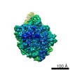
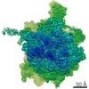
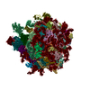
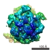
| Title | Subtomogram average of a part of the rabies lyssavirus ribonucleoprotein | |||||||||
 Map data Map data | ||||||||||
 Sample Sample |
| |||||||||
| Biological species |  Rabies virus Rabies virus | |||||||||
| Method | subtomogram averaging / cryo EM / Resolution: 15.0 Å | |||||||||
 Authors Authors | Riedel C / Vasishtan D / Prazak V / Ghanem A / Conzelmann K-K / Ruemenapf T | |||||||||
 Citation Citation | Journal: Sci Rep / Year: 2019 Title: Cryo EM structure of the rabies virus ribonucleoprotein complex. Authors: Christiane Riedel / Daven Vasishtan / Vojtěch Pražák / Alexander Ghanem / Karl-Klaus Conzelmann / Till Rümenapf /    Abstract: Rabies virus is an important zoonotic pathogen. Its bullet shaped particle contains a helical nucleocapsid. We used cryo-electron tomography and subsequent subtomogram averaging to determine the ...Rabies virus is an important zoonotic pathogen. Its bullet shaped particle contains a helical nucleocapsid. We used cryo-electron tomography and subsequent subtomogram averaging to determine the structure of its ribonucleoprotein. The resulting electron density map allowed for confident fitting of the N-protein crystal structure, indicating that interactions between neighbouring N-proteins are only mediated by N- and C-terminal protruding subdomains (aa 1-27 and aa 355-372). Additional connecting densities, likely stabilizing the ribonucleoprotein complex, are present between neighbouring M-protein densities on the same helical turn and between M- and N-protein densities located on neighbouring helical turns, but not between M-proteins of different turns, as is observed for the related Vesicular stomatitis virus (VSV). This insight into the architecture of the rabies virus nucleocapsid highlights the surprising structural divergence of large biological assemblies even if the building blocks - here exemplified by VSV M- and N-protein - are structurally closely related. | |||||||||
| History |
|
- Structure visualization
| Movie |
 Movie viewer Movie viewer |
|---|---|
| Structure viewer | EM map: SurfViewMolmilJmol/JSmol |
| Supplemental images |

- Downloads & links
-EMDB archive
| Map data | emd_4995.map.gz | 675.4 KB | EMDB map data format | |
|---|---|---|---|---|
| Header (meta data) | emd-4995-v30.xmlemd-4995.xml | 7.6 KB 7.6 KB | Display Display | EMDB header |
| Images | 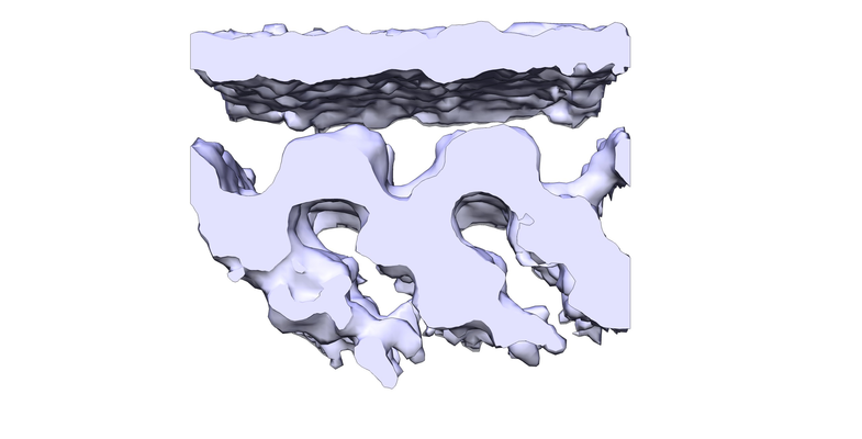 emd_4995.png emd_4995.png | 124.2 KB | ||
| Archive directory |  http://ftp.pdbj.org/pub/emdb/structures/EMD-4995ftp://ftp.pdbj.org/pub/emdb/structures/EMD-4995 http://ftp.pdbj.org/pub/emdb/structures/EMD-4995ftp://ftp.pdbj.org/pub/emdb/structures/EMD-4995 | HTTPS FTP |
-Related structure data
| Similar structure data |
|---|

-Links
| EMDB pages | EMDB (EBI/PDBe) / EMDataResource |
|---|
-Map
| File | Download / File: emd_4995.map.gz / Format: CCP4 / Size: 844.7 KB / Type: IMAGE STORED AS FLOATING POINT NUMBER (4 BYTES) | ||||||||||||||||||||||||||||||||||||||||||||||||||||||||||||||||||||
|---|---|---|---|---|---|---|---|---|---|---|---|---|---|---|---|---|---|---|---|---|---|---|---|---|---|---|---|---|---|---|---|---|---|---|---|---|---|---|---|---|---|---|---|---|---|---|---|---|---|---|---|---|---|---|---|---|---|---|---|---|---|---|---|---|---|---|---|---|---|
| Projections & slices | Image control
Images are generated by Spider. | ||||||||||||||||||||||||||||||||||||||||||||||||||||||||||||||||||||
| Voxel size | X=Y=Z: 3.352 Å | ||||||||||||||||||||||||||||||||||||||||||||||||||||||||||||||||||||
| Density |
| ||||||||||||||||||||||||||||||||||||||||||||||||||||||||||||||||||||
| Symmetry | Space group: 1 | ||||||||||||||||||||||||||||||||||||||||||||||||||||||||||||||||||||
| Details | EMDB XML:
CCP4 map header:
| ||||||||||||||||||||||||||||||||||||||||||||||||||||||||||||||||||||
 Z (Sec.)
 Y (Row.)
 X (Col.)


-Supplemental data
- Sample components
-Entire : Rabies virus
| Entire | Name: Rabies virus |
|---|---|
| Components |
|
-Supramolecule #1: Rabies virus
| Supramolecule | Name: Rabies virus / type: virus / ID: 1 / Parent: 0 / NCBI-ID: 11292 / Sci species name: Rabies virus / Virus type: VIRION / Virus isolate: STRAIN / Virus enveloped: Yes / Virus empty: No |
|---|---|
| Host system | Organism:  Homo sapiens (human) / Recombinant cell: HEK293T Homo sapiens (human) / Recombinant cell: HEK293T |
-Experimental details
-Structure determination
| Method | cryo EM |
|---|---|
 Processing Processing | subtomogram averaging |
| Aggregation state | particle |
-Sample preparation
| Buffer | pH: 7.4 |
|---|---|
| Vitrification | Cryogen name: ETHANE-PROPANE |
- Electron microscopy
| Microscope | FEI POLARA 300 |
|---|---|
| Image recording | Film or detector model: GATAN K2 SUMMIT (4k x 4k) / Average electron dose: 2.5 e/Å2 |
| Electron beam | Acceleration voltage: 300 kV / Electron source:  FIELD EMISSION GUN FIELD EMISSION GUN |
| Electron optics | Illumination mode: FLOOD BEAM / Imaging mode: BRIGHT FIELD |
| Experimental equipment |  Model: Tecnai Polara / Image courtesy: FEI Company |
-Image processing
| Final reconstruction | Applied symmetry - Point group: C1 (asymmetric) / Resolution.type: BY AUTHOR / Resolution: 15.0 Å / Resolution method: FSC 0.143 CUT-OFF / Number subtomograms used: 7079 |
|---|---|
| Extraction | Number tomograms: 10 / Number images used: 7079 |
| Final angle assignment | Type: NOT APPLICABLE |
