Movie
Movie Controller

[English] 日本語
 Yorodumi
- EMDB-49797: Structure of MurJ in complex with single gene lysis protein from ... -
+ Open data

- Basic information
| Entry |  | |||||||||
|---|---|---|---|---|---|---|---|---|---|---|
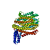
| Title | Structure of MurJ in complex with single gene lysis protein from phage PP7 | |||||||||
 Map data Map data | ||||||||||
 Sample Sample |
| |||||||||
 Keywords Keywords | lipid II flippase / phage single gene lysis proteins / peptidoglycan biosynthesis / TRANSPORT PROTEIN | |||||||||
| Function / homology |  Function and homology information Function and homology informationglycolipid translocation / lipid-linked peptidoglycan transport / lipid-linked peptidoglycan transporter activity / division septum / lipid translocation / cardiolipin binding / peptidoglycan biosynthetic process / cell wall organization / regulation of cell shape / plasma membrane Similarity search - Function | |||||||||
| Biological species |   Pseudomonas phage PP7 (virus) Pseudomonas phage PP7 (virus) | |||||||||
| Method | single particle reconstruction / cryo EM / Resolution: 3.7 Å | |||||||||
 Authors Authors | Li YE / Clemons WM | |||||||||
| Funding support |  United States, 2 items United States, 2 items
| |||||||||
 Citation Citation | Journal: Nature / Year: 2026 Title: Convergent MurJ flippase inhibition by phage lysis proteins. Authors: Yancheng E Li / S Francesca Antillon / Grace F Baron / Karthik Chamakura / Ry Young / William M Clemons / Abstract: Antimicrobial drug resistance poses a global health challenge that necessitates the identification of new druggable targets. The essential lipid II flippase MurJ is a promising yet underexplored ...Antimicrobial drug resistance poses a global health challenge that necessitates the identification of new druggable targets. The essential lipid II flippase MurJ is a promising yet underexplored antimicrobial target in bacterial cell wall biosynthesis. The only known inhibitors of Gram-negative (diderm) MurJ are the single-gene lysis proteins (Sgls) from the lytic single-strand RNA phages M (Sgl) and PP7 (Sgl). Sgl and Sgl have distinct evolutionary origins and share no sequence similarity. Here we describe a common mechanism of MurJ inhibition by these phage-encoded Sgls. We determined the structures of MurJ-bound Sgl and Sgl and discovered a third distinct MurJ-targeting Sgl from the predicted phage Changjiang3 (Sgl) that we also characterized structurally. Our findings demonstrate that all three Sgls evolved convergently to trap MurJ in a periplasm-open conformation through a common MurJ interface, revealing a pathway for drug design. | |||||||||
| History |
|
- Structure visualization
| Supplemental images |
|---|

- Downloads & links
-EMDB archive
| Map data | emd_49797.map.gz | 483.5 MB | EMDB map data format | |
|---|---|---|---|---|
| Header (meta data) | emd-49797-v30.xmlemd-49797.xml | 30 KB 30 KB | Display Display | EMDB header |
| FSC (resolution estimation) | emd_49797_fsc.xml | 17 KB | Display | FSC data file |
| Images |  emd_49797.png emd_49797.png | 67.8 KB | ||
| Masks | emd_49797_msk_1.map | 512 MB | Mask map | |
| Filedesc metadata | emd-49797.cif.gz | 7.2 KB | ||
| Others | emd_49797_additional_1.map.gzemd_49797_half_map_1.map.gzemd_49797_half_map_2.map.gz | 252.8 MB 474.8 MB 474.9 MB | ||
| Archive directory |  http://ftp.pdbj.org/pub/emdb/structures/EMD-49797ftp://ftp.pdbj.org/pub/emdb/structures/EMD-49797 http://ftp.pdbj.org/pub/emdb/structures/EMD-49797ftp://ftp.pdbj.org/pub/emdb/structures/EMD-49797 | HTTPS FTP |
-Related structure data
| Related structure data |  9nu5MC  9nu4C  9nu8C C: citing same article ( M: atomic model generated by this map |
|---|---|
| Similar structure data |


-Links
| EMDB pages | EMDB (EBI/PDBe) / EMDataResource |
|---|
-Map
| File | Download / File: emd_49797.map.gz / Format: CCP4 / Size: 512 MB / Type: IMAGE STORED AS FLOATING POINT NUMBER (4 BYTES) | ||||||||||||||||||||||||||||||||||||
|---|---|---|---|---|---|---|---|---|---|---|---|---|---|---|---|---|---|---|---|---|---|---|---|---|---|---|---|---|---|---|---|---|---|---|---|---|---|
| Projections & slices | Image control
Images are generated by Spider. | ||||||||||||||||||||||||||||||||||||
| Voxel size | X=Y=Z: 0.65 Å | ||||||||||||||||||||||||||||||||||||
| Density |
| ||||||||||||||||||||||||||||||||||||
| Symmetry | Space group: 1 | ||||||||||||||||||||||||||||||||||||
| Details | EMDB XML:
|
 Z (Sec.)
 Y (Row.)
 X (Col.)


-Supplemental data
-Mask #1
| File | emd_49797_msk_1.map | ||||||||||||
|---|---|---|---|---|---|---|---|---|---|---|---|---|---|
| Projections & Slices |
| ||||||||||||
| Density Histograms |


-Additional map: #1
| File | emd_49797_additional_1.map | ||||||||||||
|---|---|---|---|---|---|---|---|---|---|---|---|---|---|
| Projections & Slices |
| ||||||||||||
| Density Histograms |


-Half map: #1
| File | emd_49797_half_map_1.map | ||||||||||||
|---|---|---|---|---|---|---|---|---|---|---|---|---|---|
| Projections & Slices |
| ||||||||||||
| Density Histograms |


-Half map: #2
| File | emd_49797_half_map_2.map | ||||||||||||
|---|---|---|---|---|---|---|---|---|---|---|---|---|---|
| Projections & Slices |
| ||||||||||||
| Density Histograms |


- Sample components
-Entire : MurJ in complex with single gene lysis protein from phage PP7
| Entire | Name: MurJ in complex with single gene lysis protein from phage PP7 |
|---|---|
| Components |
|
-Supramolecule #1: MurJ in complex with single gene lysis protein from phage PP7
| Supramolecule | Name: MurJ in complex with single gene lysis protein from phage PP7 type: complex / ID: 1 / Parent: 0 / Macromolecule list: all |
|---|---|
| Molecular weight | Theoretical: 6 KDa |
-Supramolecule #2: soluble cytochrome b562, lipid II flippase MurJ fusion
| Supramolecule | Name: soluble cytochrome b562, lipid II flippase MurJ fusion type: complex / ID: 2 / Parent: 1 / Macromolecule list: #1 |
|---|---|
| Source (natural) | Organism: |
-Supramolecule #3: single gene lysis protein from phage PP7
| Supramolecule | Name: single gene lysis protein from phage PP7 / type: complex / ID: 3 / Parent: 1 / Macromolecule list: #2 |
|---|---|
| Source (natural) | Organism: Pseudomonas phage PP7 (virus) |
-Macromolecule #1: Lipid II flippase MurJ
| Macromolecule | Name: Lipid II flippase MurJ / type: protein_or_peptide / ID: 1 / Number of copies: 1 / Enantiomer: LEVO |
|---|---|
| Source (natural) | Organism: |
| Molecular weight | Theoretical: 66.66868 KDa |
| Recombinant expression | Organism: |
| Sequence | String: MADLEDNWET LNDNLKVIEK ADNAAQVKDA LTKMRAAALD AQKATPPKLE DKSPDSPEMK DFRHGFDILV GQIDDALKLA NEGKVKEAQ AAAEQLKTTR NAYIQKYLLF SLAAVSIATM FSRVLGFARD AIVARIFGAG MATDAFFVAF KLPNLLRRIF A EGAFSQAF ...String: MADLEDNWET LNDNLKVIEK ADNAAQVKDA LTKMRAAALD AQKATPPKLE DKSPDSPEMK DFRHGFDILV GQIDDALKLA NEGKVKEAQ AAAEQLKTTR NAYIQKYLLF SLAAVSIATM FSRVLGFARD AIVARIFGAG MATDAFFVAF KLPNLLRRIF A EGAFSQAF VPILAEYKSK QGEDATRVFV SYVSGLLTLA LAVVTVAGML AAPWVIMVTA PGFADTADKF ALTSQLLKIT FP YILLISL ASLVGAILNT WNRFSIPAFA PTLLNISMIG FALFAAPYFN PPVLALAWAV TVGGVLQLVY QLPHLKKIGM LVL PRINFH DAGAMRVVKQ MGPAILGVSV SQISLIINTI FASFLASGSV SWMYYADRLM EFPSGVLGVA LGTILLPSLS KSFA SGNHD EYNRLMDWGL RLCFLLALPS AVALGILSGP LTVSLFQYGK FTAFDALMTQ RALIAYSVGL IGLIVVKVLA PGFYS RQDI KTPVKIAIVT LILTQLMNLA FIGPLKHAGL SLSIGLAACL NASLLYWQLR KQKIFTPQPG WMAFLLRLVV AVLVMS GVL LGMLHIMPEW SLGTMPWRLL RLMAVVLAGI AAYFAALAVL GSAWSHPQFE K UniProtKB: Lipid II flippase MurJ |
-Macromolecule #2: Lysis protein
| Macromolecule | Name: Lysis protein / type: protein_or_peptide / ID: 2 / Number of copies: 1 / Enantiomer: LEVO |
|---|---|
| Source (natural) | Organism: Pseudomonas phage PP7 (virus) |
| Molecular weight | Theoretical: 6.058651 KDa |
| Recombinant expression | Organism: |
| Sequence | String: MGHHHHHHGS GSGSGSGSGS GSGSGSGSGS SKPLVALAYV TLYLLASVFL SQLAYPIGSW AV UniProtKB: Lysis protein |
-Experimental details
-Structure determination
| Method | cryo EM |
|---|---|
 Processing Processing | single particle reconstruction |
| Aggregation state | particle |
-Sample preparation
| Concentration | 4 mg/mL | ||||||||||||||||||
|---|---|---|---|---|---|---|---|---|---|---|---|---|---|---|---|---|---|---|---|
| Buffer | pH: 8 Component:
| ||||||||||||||||||
| Grid | Model: Quantifoil R1.2/1.3 / Material: COPPER / Mesh: 300 / Support film - Material: CARBON / Support film - topology: HOLEY / Pretreatment - Type: GLOW DISCHARGE / Pretreatment - Time: 60 sec. | ||||||||||||||||||
| Vitrification | Cryogen name: ETHANE / Chamber humidity: 100 % / Chamber temperature: 277.15 K / Instrument: FEI VITROBOT MARK IV |
- Electron microscopy
| Microscope | TFS KRIOS |
|---|---|
| Image recording | Film or detector model: GATAN K3 (6k x 4k) / Average electron dose: 70.0 e/Å2 |
| Electron beam | Acceleration voltage: 300 kV / Electron source:  FIELD EMISSION GUN FIELD EMISSION GUN |
| Electron optics | Illumination mode: OTHER / Imaging mode: BRIGHT FIELD / Nominal defocus max: 3.0 µm / Nominal defocus min: 1.0 µm / Nominal magnification: 130000 |
| Sample stage | Specimen holder model: FEI TITAN KRIOS AUTOGRID HOLDER / Cooling holder cryogen: NITROGEN |
| Experimental equipment |  Model: Titan Krios / Image courtesy: FEI Company |