 Movie
Movie Controller

[English] 日本語
 Yorodumi
- EMDB-49618: Human sweet taste receptor (TAS1R2 + TAS1R3) VFT domains bound to... -
+ Open data

- Basic information
| Entry |  | |||||||||
|---|---|---|---|---|---|---|---|---|---|---|
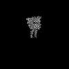
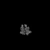
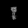
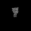
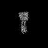
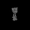
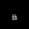
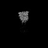
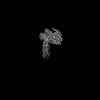
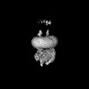
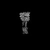
| Title | Human sweet taste receptor (TAS1R2 + TAS1R3) VFT domains bound to sucralose | |||||||||
 Map data Map data | ||||||||||
 Sample Sample |
| |||||||||
 Keywords Keywords | Sweet / taste / receptor / GPCR / TAS1R2 / TAS1R3 / T1R2 / T1R3 / MEMBRANE PROTEIN | |||||||||
| Function / homology |  Function and homology information Function and homology informationsweet taste receptor complex / detection of chemical stimulus involved in sensory perception of sweet taste / sweet taste receptor activity / sensory perception of umami taste / taste receptor activity / sensory perception of sweet taste / Class C/3 (Metabotropic glutamate/pheromone receptors) / positive regulation of cytokinesis / G protein-coupled receptor activity / Sensory perception of sweet, bitter, and umami (glutamate) taste ...sweet taste receptor complex / detection of chemical stimulus involved in sensory perception of sweet taste / sweet taste receptor activity / sensory perception of umami taste / taste receptor activity / sensory perception of sweet taste / Class C/3 (Metabotropic glutamate/pheromone receptors) / positive regulation of cytokinesis / G protein-coupled receptor activity / Sensory perception of sweet, bitter, and umami (glutamate) taste / G alpha (i) signalling events / signaling receptor complex / G protein-coupled receptor signaling pathway / Golgi apparatus / membrane / plasma membrane Similarity search - Function | |||||||||
| Biological species |  Homo sapiens (human) Homo sapiens (human) | |||||||||
| Method | single particle reconstruction / cryo EM / Resolution: 3.3 Å | |||||||||
 Authors Authors | Juen Z / Lu Z / Yu R / Chang AN / Wang B / Fitzpatrick AWP / Zuker CS | |||||||||
| Funding support |  United States, 1 items United States, 1 items
| |||||||||
 Citation Citation | Journal: Cell / Year: 2025 Title: The structure of human sweetness. Authors: Zhang Juen / Zhengyuan Lu / Ruihuan Yu / Andrew N Chang / Brian Wang / Anthony W P Fitzpatrick / Charles S Zuker / Abstract: In humans, the detection and ultimately the perception of sweetness begin in the oral cavity, where taste receptor cells (TRCs) dedicated to sweet-sensing interact with sugars, artificial sweeteners, ...In humans, the detection and ultimately the perception of sweetness begin in the oral cavity, where taste receptor cells (TRCs) dedicated to sweet-sensing interact with sugars, artificial sweeteners, and other sweet-tasting chemicals. Human sweet TRCs express on their cell surface a sweet receptor that initiates the cascade of signaling events responsible for our strong attraction to sweet stimuli. Here, we describe the cryo-electron microscopy (cryo-EM) structure of the human sweet receptor bound to two of the most widely used artificial sweeteners-sucralose and aspartame. Our results reveal the structural basis for sweet detection, provide insights into how a single receptor mediates all our responses to such a wide range of sweet-tasting compounds, and open up unique possibilities for designing a generation of taste modulators informed by the structure of the human receptor. | |||||||||
| History |
|
- Structure visualization
| Supplemental images |
|---|

- Downloads & links
-EMDB archive
| Map data | emd_49618.map.gz | 307 MB | EMDB map data format | |
|---|---|---|---|---|
| Header (meta data) | emd-49618-v30.xmlemd-49618.xml | 18.1 KB 18.1 KB | Display Display | EMDB header |
| Images |  emd_49618.png emd_49618.png | 66 KB | ||
| Masks | emd_49618_msk_1.map | 325 MB | Mask map | |
| Filedesc metadata | emd-49618.cif.gz | 6.5 KB | ||
| Others | emd_49618_half_map_1.map.gzemd_49618_half_map_2.map.gz | 301.1 MB 301.1 MB | ||
| Archive directory |  http://ftp.pdbj.org/pub/emdb/structures/EMD-49618ftp://ftp.pdbj.org/pub/emdb/structures/EMD-49618 http://ftp.pdbj.org/pub/emdb/structures/EMD-49618ftp://ftp.pdbj.org/pub/emdb/structures/EMD-49618 | HTTPS FTP |
-Related structure data
| Related structure data |  9novMC  9norC  9nosC  9notC 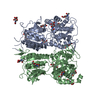 9nouC 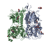 9nowC 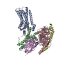 9noxC 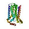 9noyC 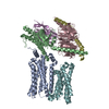 9o38C M: atomic model generated by this map C: citing same article ( |
|---|---|
| Similar structure data |


-Links
| EMDB pages | EMDB (EBI/PDBe) / EMDataResource |
|---|---|
| Related items in Molecule of the Month |


-Map
| File | Download / File: emd_49618.map.gz / Format: CCP4 / Size: 325 MB / Type: IMAGE STORED AS FLOATING POINT NUMBER (4 BYTES) | ||||||||||||||||||||||||||||||||||||
|---|---|---|---|---|---|---|---|---|---|---|---|---|---|---|---|---|---|---|---|---|---|---|---|---|---|---|---|---|---|---|---|---|---|---|---|---|---|
| Projections & slices | Image control
Images are generated by Spider. | ||||||||||||||||||||||||||||||||||||
| Voxel size | X=Y=Z: 0.823 Å | ||||||||||||||||||||||||||||||||||||
| Density |
| ||||||||||||||||||||||||||||||||||||
| Symmetry | Space group: 1 | ||||||||||||||||||||||||||||||||||||
| Details | EMDB XML:
|
 Z (Sec.)
 Y (Row.)
 X (Col.)


-Supplemental data
-Mask #1
| File | emd_49618_msk_1.map | ||||||||||||
|---|---|---|---|---|---|---|---|---|---|---|---|---|---|
| Projections & Slices |
| ||||||||||||
| Density Histograms |


-Half map: #2
| File | emd_49618_half_map_1.map | ||||||||||||
|---|---|---|---|---|---|---|---|---|---|---|---|---|---|
| Projections & Slices |
| ||||||||||||
| Density Histograms |


-Half map: #1
| File | emd_49618_half_map_2.map | ||||||||||||
|---|---|---|---|---|---|---|---|---|---|---|---|---|---|
| Projections & Slices |
| ||||||||||||
| Density Histograms |


- Sample components
-Entire : Human sweet taste receptor (TAS1R2 + TAS1R3) VFT domains bound to...
| Entire | Name: Human sweet taste receptor (TAS1R2 + TAS1R3) VFT domains bound to sucralose |
|---|---|
| Components |
|
-Supramolecule #1: Human sweet taste receptor (TAS1R2 + TAS1R3) VFT domains bound to...
| Supramolecule | Name: Human sweet taste receptor (TAS1R2 + TAS1R3) VFT domains bound to sucralose type: complex / ID: 1 / Parent: 0 / Macromolecule list: #1-#2 |
|---|---|
| Source (natural) | Organism: Homo sapiens (human) |
-Macromolecule #1: Taste receptor type 1 member 3
| Macromolecule | Name: Taste receptor type 1 member 3 / type: protein_or_peptide / ID: 1 / Number of copies: 1 / Enantiomer: LEVO |
|---|---|
| Source (natural) | Organism: Homo sapiens (human) |
| Molecular weight | Theoretical: 58.379551 KDa |
| Recombinant expression | Organism: Homo sapiens (human) |
| Sequence | String: MKTIIALSYI FCLVFADYKD DDDKAAAAPL CLSQQLRMKG DYVLGGLFPL GEAEEAGLRS RTRPSSPVCT RFSSNGLLWA LAMKMAVEE INNKSDLLPG LRLGYDLFDT CSEPVVAMKP SLMFLAKAGS RDIAAYCNYT QYQPRVLAVI GPHSSELAMV T GKFFSFFL ...String: MKTIIALSYI FCLVFADYKD DDDKAAAAPL CLSQQLRMKG DYVLGGLFPL GEAEEAGLRS RTRPSSPVCT RFSSNGLLWA LAMKMAVEE INNKSDLLPG LRLGYDLFDT CSEPVVAMKP SLMFLAKAGS RDIAAYCNYT QYQPRVLAVI GPHSSELAMV T GKFFSFFL MPQVSYGASM ELLSARETFP SFFRTVPSDR VQLTAAAELL QEFGWNWVAA LGSDDEYGRQ GLSIFSALAA AR GICIAHE GLVPLPRADD SRLGKVQDVL HQVNQSSVQV VLLFASVHAA HALFNYSISS RLSPKVWVAS EAWLTSDLVM GLP GMAQMG TVLGFLQRGA QLHEFPQYVK THLALATDPA FCSALGEREQ GLEEDVVGQR CPQCDCITLQ NVSAGLNHHQ TFSV YAAVY SVAQALHNTL QCNASGCPAQ DPVKPWQLLE NMYNLTFHVG GLPLRFDSSG NVDMEYDLKL WVWQGSVPRL HDVGR FNGS LRTERLKIRW HTSDNQKPVS RCSRQCQEGQ VRRVKGFHSC CYDC UniProtKB: Taste receptor type 1 member 3 |
-Macromolecule #2: Taste receptor type 1 member 2
| Macromolecule | Name: Taste receptor type 1 member 2 / type: protein_or_peptide / ID: 2 / Number of copies: 1 / Enantiomer: LEVO |
|---|---|
| Source (natural) | Organism: Homo sapiens (human) |
| Molecular weight | Theoretical: 60.070512 KDa |
| Recombinant expression | Organism: Homo sapiens (human) |
| Sequence | String: MKTIIALSYI FCLVFAYPYD VPDYAAAAEP AENSDFYLPG DYLLGGLFSL HANMKGIVHL NFLQVPMCKE YEVKVIGYNL MQAMRFAVE EINNDSSLLP GVLLGYEIVD VCYISNNVQP VLYFLAHEDN LLPIQEDYSN YISRVVAVIG PDNSESVMTV A NFLSLFLL ...String: MKTIIALSYI FCLVFAYPYD VPDYAAAAEP AENSDFYLPG DYLLGGLFSL HANMKGIVHL NFLQVPMCKE YEVKVIGYNL MQAMRFAVE EINNDSSLLP GVLLGYEIVD VCYISNNVQP VLYFLAHEDN LLPIQEDYSN YISRVVAVIG PDNSESVMTV A NFLSLFLL PQITYSAISD ELRDKVRFPA LLRTTPSADH HIEAMVQLML HFRWNWIIVL VSSDTYGRDN GQLLGERVAR RD ICIAFQE TLPTLQPNQN MTSEERQRLV TIVDKLQQST ARVVVVFSPD LTLYHFFNEV LRQNFTGAVW IASESWAIDP VLH NLTELR HLGTFLGITI QSVPIPGFSE FREWGPQAGP PPLSRTSQSY TCNQECDNCL NATLSFNTIL RLSGERVVYS VYSA VYAVA HALHSLLGCD KSTCTKRVVY PWQLLEEIWK VNFTLLDHQI FFDPQGDVAL HLEIVQWQWD RSQNPFQSVA SYYPL QRQL KNIQDISWHT INNTIPMSMC SKRCQSGQKK KPVGIHVCCF ECI UniProtKB: Taste receptor type 1 member 2 |
-Macromolecule #4: 2-acetamido-2-deoxy-beta-D-glucopyranose
| Macromolecule | Name: 2-acetamido-2-deoxy-beta-D-glucopyranose / type: ligand / ID: 4 / Number of copies: 15 / Formula: NAG |
|---|---|
| Molecular weight | Theoretical: 221.208 Da |
| Chemical component information |  ChemComp-NAG: |
-Experimental details
-Structure determination
| Method | cryo EM |
|---|---|
 Processing Processing | single particle reconstruction |
| Aggregation state | particle |
-Sample preparation
| Buffer | pH: 7.5 |
|---|---|
| Vitrification | Cryogen name: ETHANE |
- Electron microscopy
| Microscope | TFS KRIOS |
|---|---|
| Image recording | Film or detector model: GATAN K3 BIOQUANTUM (6k x 4k) / Average electron dose: 59.0 e/Å2 |
| Electron beam | Acceleration voltage: 300 kV / Electron source:  FIELD EMISSION GUN FIELD EMISSION GUN |
| Electron optics | Illumination mode: FLOOD BEAM / Imaging mode: BRIGHT FIELD / Nominal defocus max: 2.0 µm / Nominal defocus min: 1.2 µm |
| Experimental equipment |  Model: Titan Krios / Image courtesy: FEI Company |
