 Movie
Movie Controller


+ Open data

- Basic information
| Entry |  | |||||||||||||||
|---|---|---|---|---|---|---|---|---|---|---|---|---|---|---|---|---|
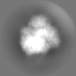
| Title | Structure of cytoplasmic 80S ribosome from S. cerevisiae | |||||||||||||||
 Map data Map data | Subtomogram average of 80S S. cerevisiae ribosome | |||||||||||||||
 Sample Sample |
| |||||||||||||||
 Keywords Keywords | ribosome / cytoplasm | |||||||||||||||
| Biological species |  | |||||||||||||||
| Method | subtomogram averaging / cryo EM / Resolution: 8.0 Å | |||||||||||||||
 Authors Authors | Chang Y / Barad BA / Rahmani H / Zid BM / Grotjahn DA | |||||||||||||||
| Funding support |  United States, 4 items United States, 4 items
| |||||||||||||||
 Citation Citation | Journal: bioRxiv / Year: 2024 Title: Cytoplasmic ribosomes on mitochondria alter the local membrane environment for protein import. Authors: Ya-Ting Chang / Benjamin A Barad / Hamidreza Rahmani / Brian M Zid / Danielle A Grotjahn / Abstract: Most of the mitochondria proteome is nuclear-encoded, synthesized by cytoplasmic ribosomes, and targeted to mitochondria post-translationally. However, a subset of mitochondrial-targeted proteins is ...Most of the mitochondria proteome is nuclear-encoded, synthesized by cytoplasmic ribosomes, and targeted to mitochondria post-translationally. However, a subset of mitochondrial-targeted proteins is imported co-translationally, although the molecular mechanisms governing this process remain unclear. We employ cellular cryo-electron tomography to visualize interactions between cytoplasmic ribosomes and mitochondria in . We use surface morphometrics tools to identify a subset of ribosomes optimally oriented on mitochondrial membranes for protein import. This allows us to establish the first subtomogram average structure of a cytoplasmic ribosome on the surface of the mitochondria in the native cellular context, which showed three distinct connections with the outer mitochondrial membrane surrounding the peptide exit tunnel. Further, this analysis demonstrated that cytoplasmic ribosomes primed for mitochondrial protein import cluster on the outer mitochondrial membrane at sites of local constrictions of the outer and inner mitochondrial membrane. Overall, our study reveals the architecture and the spatial organization of cytoplasmic ribosomes at the mitochondrial surface, providing a native cellular context to define the mechanisms that mediate efficient mitochondrial co-translational protein import. | |||||||||||||||
| History |
|
- Structure visualization
| Supplemental images |
|---|

- Downloads & links
-EMDB archive
| Map data | emd_48752.map.gz | 12.4 MB |  EMDB map data format EMDB map data format | |
|---|---|---|---|---|
| Header (meta data) | emd-48752-v30.xmlemd-48752.xml | 17.3 KB 17.3 KB | Display Display | EMDB header |
| FSC (resolution estimation) | emd_48752_fsc.xmlemd_48752_fsc_2.xml | 6.2 KB 7.1 KB | Display Display | FSC data file |
| Images |  emd_48752.png emd_48752.png | 59.5 KB | ||
| Filedesc metadata | emd-48752.cif.gz | 4.8 KB | ||
| Others | emd_48752_half_map_1.map.gzemd_48752_half_map_2.map.gz | 10.1 MB 10.2 MB | ||
| Archive directory |  http://ftp.pdbj.org/pub/emdb/structures/EMD-48752ftp://ftp.pdbj.org/pub/emdb/structures/EMD-48752 http://ftp.pdbj.org/pub/emdb/structures/EMD-48752ftp://ftp.pdbj.org/pub/emdb/structures/EMD-48752 | HTTPS FTP |
-Related structure data

-Links
| EMDB pages | EMDB (EBI/PDBe) / EMDataResource |
|---|
-Map
| File | Download / File: emd_48752.map.gz / Format: CCP4 / Size: 13.4 MB / Type: IMAGE STORED AS FLOATING POINT NUMBER (4 BYTES) | ||||||||||||||||||||||||||||||||||||
|---|---|---|---|---|---|---|---|---|---|---|---|---|---|---|---|---|---|---|---|---|---|---|---|---|---|---|---|---|---|---|---|---|---|---|---|---|---|
| Annotation | Subtomogram average of 80S S. cerevisiae ribosome | ||||||||||||||||||||||||||||||||||||
| Projections & slices | Image control
Images are generated by Spider. | ||||||||||||||||||||||||||||||||||||
| Voxel size | X=Y=Z: 3.33 Å | ||||||||||||||||||||||||||||||||||||
| Density |
| ||||||||||||||||||||||||||||||||||||
| Symmetry | Space group: 1 | ||||||||||||||||||||||||||||||||||||
| Details | EMDB XML:
|
 Z (Sec.)
 Y (Row.)
 X (Col.)


-Supplemental data
-Half map: #2
| File | emd_48752_half_map_1.map | ||||||||||||
|---|---|---|---|---|---|---|---|---|---|---|---|---|---|
| Projections & Slices |
| ||||||||||||
| Density Histograms |


-Half map: #1
| File | emd_48752_half_map_2.map | ||||||||||||
|---|---|---|---|---|---|---|---|---|---|---|---|---|---|
| Projections & Slices |
| ||||||||||||
| Density Histograms |


- Sample components
-Entire : Cytoplasmic 80S ribosome from S. cerevisiae
| Entire | Name: Cytoplasmic 80S ribosome from S. cerevisiae |
|---|---|
| Components |
|
-Supramolecule #1: Cytoplasmic 80S ribosome from S. cerevisiae
| Supramolecule | Name: Cytoplasmic 80S ribosome from S. cerevisiae / type: complex / ID: 1 / Parent: 0 |
|---|---|
| Source (natural) | Organism: |
-Experimental details
-Structure determination
| Method | cryo EM |
|---|---|
 Processing Processing | subtomogram averaging |
| Aggregation state | cell |
-Sample preparation
| Buffer | pH: 6.5 |
|---|---|
| Grid | Model: Quantifoil R1/4 / Material: GOLD / Mesh: 200 / Support film - Material: CARBON / Support film - topology: HOLEY / Pretreatment - Type: GLOW DISCHARGE |
| Vitrification | Cryogen name: ETHANE-PROPANE / Chamber humidity: 100 % / Chamber temperature: 303.15 K / Instrument: FEI VITROBOT MARK IV |
- Electron microscopy
| Microscope | TFS KRIOS |
|---|---|
| Specialist optics | Energy filter - Name: GIF Bioquantum / Energy filter - Slit width: 20 eV |
| Image recording | Film or detector model: GATAN K3 BIOQUANTUM (6k x 4k) / Digitization - Dimensions - Width: 5760 pixel / Digitization - Dimensions - Height: 4092 pixel / Average electron dose: 3.0 e/Å2 |
| Electron beam | Acceleration voltage: 300 kV / Electron source:  FIELD EMISSION GUN FIELD EMISSION GUN |
| Electron optics | C2 aperture diameter: 70.0 µm / Illumination mode: FLOOD BEAM / Imaging mode: BRIGHT FIELD / Nominal defocus max: 6.0 µm / Nominal defocus min: 4.0 µm / Nominal magnification: 53000 |
| Sample stage | Specimen holder model: FEI TITAN KRIOS AUTOGRID HOLDER / Cooling holder cryogen: NITROGEN |
| Experimental equipment |  Model: Titan Krios / Image courtesy: FEI Company |
-Image processing
| Final reconstruction | Applied symmetry - Point group: C1 (asymmetric) / Resolution.type: BY AUTHOR / Resolution: 8.0 Å / Resolution method: FSC 0.143 CUT-OFF / Software - Name: RELION Software - details: M was used for post-processing and final refinement Number subtomograms used: 35784 | ||||||
|---|---|---|---|---|---|---|---|
| Extraction | Number tomograms: 91 / Number images used: 35784 Software:
| ||||||
| Final angle assignment | Type: MAXIMUM LIKELIHOOD / Software - Name: RELION (ver. 4.0) / Software - details: Relion + M | ||||||
| FSC plot (resolution estimation) |  |
