National Institutes of Health/National Institute Of Allergy and Infectious Diseases (NIH/NIAID)
75N93019C00051
United States
Citation
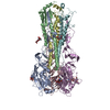
Journal: Nat Commun / Year: 2025 Title: Human monoclonal antibodies that target clade 2.3.4.4b H5N1 hemagglutinin. Authors: Garazi Peña Alzua / André Nicolás León / Temima Yellin / Disha Bhavsar / Madhumathi Loganathan / Kaitlyn Bushfield / Philip J M Brouwer / Alesandra J Rodriguez / Trushar Jeevan / Richard ...Authors: Garazi Peña Alzua / André Nicolás León / Temima Yellin / Disha Bhavsar / Madhumathi Loganathan / Kaitlyn Bushfield / Philip J M Brouwer / Alesandra J Rodriguez / Trushar Jeevan / Richard Webby / Christine Marizzi / Julianna Han / Andrew B Ward / J Andrew Duty / Florian Krammer / Abstract: The highly pathogenic avian influenza H5N1 virus clade 2.3.4.4b has been spreading globally since 2022, causing mortality and morbidity in domestic and wild birds, as well as in mammals, which ...The highly pathogenic avian influenza H5N1 virus clade 2.3.4.4b has been spreading globally since 2022, causing mortality and morbidity in domestic and wild birds, as well as in mammals, which underscores its potential to cause a pandemic. Here, we generate a panel of anti-hemagglutinin (HA) human monoclonal antibodies (mAbs) against the H5 protein of clade 2.3.4.4b. To develop human chimeric antibodies, H2L2 Harbor Mice®, which express human immunoglobulin germline genes, were immunized with H5 and N1 recombinant proteins from A/mallard/New York/22-008760-007- original/2022 H5N1 virus. Through hybridoma technology, sixteen fully human mAbs are generated, most of which show cross-reactivity against H5 proteins from different clade 2.3.4.4 virus variants. Fourteen out of the sixteen mAbs neutralize the virus in vitro. The mAbs with the strongest hemagglutination inhibition activity also demonstrate greater neutralizing capacity and show increased protective effects in vivo when administered prophylactically or therapeutically in a murine H5N1 challenge model. Using cryo-electron microscopy, we identify a cross-clonotype conserved motif that bound a hydrophobic groove on the head domain of H5 HA. Akin to mAbs against severe acute respiratory syndrome coronavirus 2 during the coronavirus 2019 pandemic, these mAbs could serve as treatments in case of a widespread H5N1 epidemic or pandemic.
In the structure databanks used in Yorodumi, some data are registered as the other names, "COVID-19 virus" and "2019-nCoV". Here are the details of the virus and the list of structure data.
Jan 31, 2019. EMDB accession codes are about to change! (news from PDBe EMDB page)
EMDB accession codes are about to change! (news from PDBe EMDB page)
The allocation of 4 digits for EMDB accession codes will soon come to an end. Whilst these codes will remain in use, new EMDB accession codes will include an additional digit and will expand incrementally as the available range of codes is exhausted. The current 4-digit format prefixed with “EMD-” (i.e. EMD-XXXX) will advance to a 5-digit format (i.e. EMD-XXXXX), and so on. It is currently estimated that the 4-digit codes will be depleted around Spring 2019, at which point the 5-digit format will come into force.
The EM Navigator/Yorodumi systems omit the EMD- prefix.
Related info.:Q: What is EMD? / ID/Accession-code notation in Yorodumi/EM Navigator
Yorodumi is a browser for structure data from EMDB, PDB, SASBDB, etc.
This page is also the successor to EM Navigator detail page, and also detail information page/front-end page for Omokage search.
The word "yorodu" (or yorozu) is an old Japanese word meaning "ten thousand". "mi" (miru) is to see.
Related info.:EMDB / PDB / SASBDB / Comparison of 3 databanks / Yorodumi Search / Aug 31, 2016. New EM Navigator & Yorodumi / Yorodumi Papers / Jmol/JSmol / Function and homology information / Changes in new EM Navigator and Yorodumi
 Movie
Movie Controller

 Yorodumi
 Open data
 Basic information
 Map data
 Sample
 Keywords
 Homo sapiens (human)
 Authors
 United States, 1 items
  Citation
 Structure visualization
 Downloads & links
 EMDB map data format
 emd_48688.png
 http://ftp.pdbj.org/pub/emdb/structures/EMD-48688


 Z (Sec.)
 Y (Row.)
 X (Col.)


 Sample components
 Processing
 Electron microscopy