Movie
Movie Controller

[English] 日本語
 Yorodumi
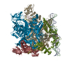
- EMDB-48580: de novo SigN RNA polymerase transcription initiation intermediate... -
+ Open data

- Basic information
| Entry |  | |||||||||
|---|---|---|---|---|---|---|---|---|---|---|
| Title | de novo SigN RNA polymerase transcription initiation intermediate with pre-catalytic bEBP state (RPi1 open ring), consensus map | |||||||||
 Map data Map data | unsharpened consensus map | |||||||||
 Sample Sample |
| |||||||||
 Keywords Keywords | sigma N / sigma 54 / ATPase / bacterial enhancer binding protein / transcription initiation / intermediate / TRANSCRIPTION | |||||||||
| Biological species |  | |||||||||
| Method | single particle reconstruction / cryo EM / Resolution: 2.7 Å | |||||||||
 Authors Authors | Mueller AU / Darst SA | |||||||||
| Funding support |  United States, 2 items United States, 2 items
| |||||||||
 Citation Citation | Journal: Nat Commun / Year: 2025 Title: Real-time capture of σ transcription initiation intermediates reveals mechanism of ATPase-driven activation by limited unfolding. Authors: Andreas U Mueller / Nina Molina / B Tracy Nixon / Seth A Darst / Abstract: Bacterial σ factors bind RNA polymerase (E) to form holoenzyme (Eσ), conferring promoter specificity to E and playing a key role in transcription bubble formation. σ is unique among σ factors in ...Bacterial σ factors bind RNA polymerase (E) to form holoenzyme (Eσ), conferring promoter specificity to E and playing a key role in transcription bubble formation. σ is unique among σ factors in its structure and functional mechanism, requiring activation by specialized AAA+ ATPases. Eσ forms an inactive promoter complex where the N-terminal σ region I (σ-RI) threads through a small DNA bubble. On the opposite side of the DNA, the ATPase engages σ-RI within the pore of its hexameric ring. Here, we perform kinetics-guided structural analysis of de novo formed Eσ initiation complexes and engineer a biochemical assay to measure ATPase-mediated σ-RI translocation during promoter melting. We show that the ATPase exerts mechanical action to translocate about 30 residues of σ-RI through the DNA bubble, disrupting inhibitory structures of σ to allow full transcription bubble formation. A local charge switch of σ-RI from positive to negative may help facilitate disengagement of the otherwise processive ATPase, allowing subsequent σ disentanglement from the DNA bubble. | |||||||||
| History |
|
- Structure visualization
| Supplemental images |
|---|

- Downloads & links
-EMDB archive
| Map data | emd_48580.map.gz | 172.1 MB |  EMDB map data format EMDB map data format | |
|---|---|---|---|---|
| Header (meta data) | emd-48580-v30.xmlemd-48580.xml | 17.7 KB 17.7 KB | Display Display | EMDB header |
| FSC (resolution estimation) | emd_48580_fsc.xml | 14.8 KB | Display | FSC data file |
| Images |  emd_48580.png emd_48580.png | 109 KB | ||
| Filedesc metadata | emd-48580.cif.gz | 4.6 KB | ||
| Others | emd_48580_additional_1.map.gzemd_48580_half_map_1.map.gzemd_48580_half_map_2.map.gz | 324.2 MB 318.1 MB 318.1 MB | ||
| Archive directory |  http://ftp.pdbj.org/pub/emdb/structures/EMD-48580ftp://ftp.pdbj.org/pub/emdb/structures/EMD-48580 http://ftp.pdbj.org/pub/emdb/structures/EMD-48580ftp://ftp.pdbj.org/pub/emdb/structures/EMD-48580 | HTTPS FTP |
-Validation report
| Summary document | emd_48580_validation.pdf.gz | 971.5 KB | Display | EMDB validaton report |
|---|---|---|---|---|
| Full document | emd_48580_full_validation.pdf.gz | 971.1 KB | Display | |
| Data in XML | emd_48580_validation.xml.gz | 24.2 KB | Display | |
| Data in CIF | emd_48580_validation.cif.gz | 31.5 KB | Display | |
| Arichive directory | https://ftp.pdbj.org/pub/emdb/validation_reports/EMD-48580ftp://ftp.pdbj.org/pub/emdb/validation_reports/EMD-48580 | HTTPS FTP |
-Related structure data


-Links
| EMDB pages | EMDB (EBI/PDBe) / EMDataResource |
|---|
-Map
| File | Download / File: emd_48580.map.gz / Format: CCP4 / Size: 343 MB / Type: IMAGE STORED AS FLOATING POINT NUMBER (4 BYTES) | ||||||||||||||||||||||||||||||||||||
|---|---|---|---|---|---|---|---|---|---|---|---|---|---|---|---|---|---|---|---|---|---|---|---|---|---|---|---|---|---|---|---|---|---|---|---|---|---|
| Annotation | unsharpened consensus map | ||||||||||||||||||||||||||||||||||||
| Projections & slices | Image control
Images are generated by Spider. | ||||||||||||||||||||||||||||||||||||
| Voxel size | X=Y=Z: 0.86 Å | ||||||||||||||||||||||||||||||||||||
| Density |
| ||||||||||||||||||||||||||||||||||||
| Symmetry | Space group: 1 | ||||||||||||||||||||||||||||||||||||
| Details | EMDB XML:
|
 Z (Sec.)
 Y (Row.)
 X (Col.)


-Supplemental data
-Additional map: sharpened consensus map (b-factor 61.6)
| File | emd_48580_additional_1.map | ||||||||||||
|---|---|---|---|---|---|---|---|---|---|---|---|---|---|
| Annotation | sharpened consensus map (b-factor 61.6) | ||||||||||||
| Projections & Slices |
| ||||||||||||
| Density Histograms |


-Half map: half map B
| File | emd_48580_half_map_1.map | ||||||||||||
|---|---|---|---|---|---|---|---|---|---|---|---|---|---|
| Annotation | half map B | ||||||||||||
| Projections & Slices |
| ||||||||||||
| Density Histograms |


-Half map: half map A
| File | emd_48580_half_map_2.map | ||||||||||||
|---|---|---|---|---|---|---|---|---|---|---|---|---|---|
| Annotation | half map A | ||||||||||||
| Projections & Slices |
| ||||||||||||
| Density Histograms |


- Sample components
-Entire : EsNdhsUC1+ATP
| Entire | Name: EsNdhsUC1+ATP |
|---|---|
| Components |
|
-Supramolecule #1: EsNdhsUC1+ATP
| Supramolecule | Name: EsNdhsUC1+ATP / type: complex / ID: 1 / Parent: 0 / Macromolecule list: #1-#8 Details: E = RNAP sN = Sigma N dhsU = dhsU promoter DNA C1 = NtrC1 |
|---|---|
| Source (natural) | Organism: |
| Molecular weight | Theoretical: 180 KDa |
-Experimental details
-Structure determination
| Method | cryo EM |
|---|---|
 Processing Processing | single particle reconstruction |
| Aggregation state | particle |
-Sample preparation
| Buffer | pH: 8 Details: 40 mM Tris-HCl, pH 8/RT, 200 mM KCl, 10 mM MgCl2, 1 mM DTT; fluorinated fos-choline-8 (FC8F) added to a final concentration of 1.5 mM during grid preparation |
|---|---|
| Grid | Model: C-flat-1.2/1.3 / Material: GOLD / Mesh: 400 / Support film - Material: CARBON / Support film - topology: HOLEY |
| Vitrification | Cryogen name: ETHANE / Chamber humidity: 100 % / Chamber temperature: 310 K / Instrument: FEI VITROBOT MARK IV |
- Electron microscopy
| Microscope | TFS KRIOS |
|---|---|
| Image recording | Film or detector model: GATAN K3 BIOQUANTUM (6k x 4k) / Digitization - Dimensions - Width: 11520 pixel / Digitization - Dimensions - Height: 8184 pixel / Number grids imaged: 1 / Number real images: 17199 / Average exposure time: 1.4 sec. / Average electron dose: 42.0 e/Å2 |
| Electron beam | Acceleration voltage: 300 kV / Electron source:  FIELD EMISSION GUN FIELD EMISSION GUN |
| Electron optics | Illumination mode: FLOOD BEAM / Imaging mode: BRIGHT FIELD / Nominal defocus max: 2.2 µm / Nominal defocus min: 0.8 µm |
| Sample stage | Cooling holder cryogen: NITROGEN |
| Experimental equipment |  Model: Titan Krios / Image courtesy: FEI Company |