[English] 日本語
 Yorodumi
- PDB-9msg: De novo SigN RNA polymerase transcription initiation intermediate... -
+ Open data
- Basic information
| Entry | Database: PDB / ID: 9msg | |||||||||
|---|---|---|---|---|---|---|---|---|---|---|
| Title | De novo SigN RNA polymerase transcription initiation intermediate with bound SigN-RII | |||||||||
 Components Components |
| |||||||||
 Keywords Keywords | TRANSCRIPTION / TRANSFERASE/DNA / sigma N / sigma 54 / ATPase / bacterial enhancer binding protein / transcription initiation / intermediate / TRANSFERASE-DNA complex | |||||||||
| Function / homology |  Function and homology information Function and homology informationarginine metabolic process / DNA-binding transcription activator activity / RNA polymerase complex / submerged biofilm formation / cellular response to cell envelope stress / regulation of DNA-templated transcription initiation / phosphorelay signal transduction system / sigma factor activity / bacterial-type flagellum assembly / bacterial-type RNA polymerase core enzyme binding ...arginine metabolic process / DNA-binding transcription activator activity / RNA polymerase complex / submerged biofilm formation / cellular response to cell envelope stress / regulation of DNA-templated transcription initiation / phosphorelay signal transduction system / sigma factor activity / bacterial-type flagellum assembly / bacterial-type RNA polymerase core enzyme binding / cytosolic DNA-directed RNA polymerase complex / bacterial-type flagellum-dependent cell motility / nitrate assimilation / cis-regulatory region sequence-specific DNA binding / nucleotidyltransferase activity / regulation of DNA-templated transcription elongation / transcription elongation factor complex / transcription antitermination / DNA-directed RNA polymerase complex / cell motility / DNA-templated transcription initiation / protein-DNA complex / ribonucleoside binding / DNA-directed RNA polymerase / DNA-directed RNA polymerase activity / response to heat / protein-containing complex assembly / intracellular iron ion homeostasis / protein dimerization activity / transcription cis-regulatory region binding / response to antibiotic / DNA-templated transcription / regulation of DNA-templated transcription / positive regulation of DNA-templated transcription / magnesium ion binding / DNA binding / zinc ion binding / ATP binding / metal ion binding / identical protein binding / membrane / cytoplasm / cytosol Similarity search - Function | |||||||||
| Biological species |   Aquifex aeolicus VF5 (bacteria) Aquifex aeolicus VF5 (bacteria) | |||||||||
| Method | ELECTRON MICROSCOPY / single particle reconstruction / cryo EM / Resolution: 2.7 Å | |||||||||
 Authors Authors | Mueller, A.U. / Darst, S.A. | |||||||||
| Funding support |  United States, 2items United States, 2items
| |||||||||
 Citation Citation |  Journal: Nat Commun / Year: 2025 Journal: Nat Commun / Year: 2025Title: Real-time capture of σ transcription initiation intermediates reveals mechanism of ATPase-driven activation by limited unfolding. Authors: Andreas U Mueller / Nina Molina / B Tracy Nixon / Seth A Darst /  Abstract: Bacterial σ factors bind RNA polymerase (E) to form holoenzyme (Eσ), conferring promoter specificity to E and playing a key role in transcription bubble formation. σ is unique among σ factors in ...Bacterial σ factors bind RNA polymerase (E) to form holoenzyme (Eσ), conferring promoter specificity to E and playing a key role in transcription bubble formation. σ is unique among σ factors in its structure and functional mechanism, requiring activation by specialized AAA+ ATPases. Eσ forms an inactive promoter complex where the N-terminal σ region I (σ-RI) threads through a small DNA bubble. On the opposite side of the DNA, the ATPase engages σ-RI within the pore of its hexameric ring. Here, we perform kinetics-guided structural analysis of de novo formed Eσ initiation complexes and engineer a biochemical assay to measure ATPase-mediated σ-RI translocation during promoter melting. We show that the ATPase exerts mechanical action to translocate about 30 residues of σ-RI through the DNA bubble, disrupting inhibitory structures of σ to allow full transcription bubble formation. A local charge switch of σ-RI from positive to negative may help facilitate disengagement of the otherwise processive ATPase, allowing subsequent σ disentanglement from the DNA bubble. | |||||||||
| History |
|
- Structure visualization
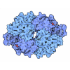
| Structure viewer | Molecule:  Molmil Molmil Jmol/JSmol Jmol/JSmol |
|---|
- Downloads & links
- Download
| PDBx/mmCIF format |  9msg.cif.gz 9msg.cif.gz | 1 MB | Display |  PDBx/mmCIF format PDBx/mmCIF format |
|---|---|---|---|---|
| PDB format |  pdb9msg.ent.gz pdb9msg.ent.gz | 859.7 KB | Display |  PDB format PDB format |
| PDBx/mmJSON format |  9msg.json.gz 9msg.json.gz | Tree view |  PDBx/mmJSON format PDBx/mmJSON format | |
| Others |  Other downloads Other downloads |
-Validation report
| Arichive directory |  https://data.pdbj.org/pub/pdb/validation_reports/ms/9msg https://data.pdbj.org/pub/pdb/validation_reports/ms/9msg ftp://data.pdbj.org/pub/pdb/validation_reports/ms/9msg ftp://data.pdbj.org/pub/pdb/validation_reports/ms/9msg | HTTPS FTP |
|---|
-Related structure data
| Related structure data |  48588MC  9mseC  9msfC  9mshC  9msjC M: map data used to model this data C: citing same article ( |
|---|---|
| Similar structure data | Similarity search - Function & homology  F&H Search F&H Search |
- Links
Links
- Assembly
| Deposited unit | 
|
|---|---|
| 1 |
|
- Components
-Protein , 2 types, 7 molecules ABCDEFM
| #1: Protein | Mass: 30731.598 Da / Num. of mol.: 6 Source method: isolated from a genetically manipulated source Details: AAA+ domain of NtrC1 (UNP residues 121-387) / Source: (gene. exp.)   Aquifex aeolicus VF5 (bacteria) / Gene: ntrC1, aq_1117 / Production host: Aquifex aeolicus VF5 (bacteria) / Gene: ntrC1, aq_1117 / Production host:  #6: Protein | | Mass: 54043.543 Da / Num. of mol.: 1 Source method: isolated from a genetically manipulated source Source: (gene. exp.)   |
|---|
-DNA-directed RNA polymerase subunit ... , 4 types, 5 molecules GHIJK
| #2: Protein | Mass: 36558.680 Da / Num. of mol.: 2 Source method: isolated from a genetically manipulated source Source: (gene. exp.)   #3: Protein | | Mass: 150820.875 Da / Num. of mol.: 1 Source method: isolated from a genetically manipulated source Source: (gene. exp.)  Gene: rpoB, groN, nitB, rif, ron, stl, stv, tabD, b3987, JW3950 Production host:  #4: Protein | | Mass: 156338.891 Da / Num. of mol.: 1 Source method: isolated from a genetically manipulated source Source: (gene. exp.)   #5: Protein | | Mass: 10249.547 Da / Num. of mol.: 1 Source method: isolated from a genetically manipulated source Source: (gene. exp.)   |
|---|
-DhsU (-60 to +30) ... , 2 types, 2 molecules UV
| #7: DNA chain | Mass: 28003.027 Da / Num. of mol.: 1 / Source method: obtained synthetically Details: Bases at the ends of the fragment (two positions on either side) were modified from the genomic sequence to G or C to stabilize the ends. Source: (synth.)   Aquifex aeolicus VF5 (bacteria) / References: GenBank: 6626248 Aquifex aeolicus VF5 (bacteria) / References: GenBank: 6626248 |
|---|---|
| #8: DNA chain | Mass: 27507.594 Da / Num. of mol.: 1 / Source method: obtained synthetically Details: Bases at the ends of the fragment (two positions on either side) were modified from the genomic sequence to G or C to stabilize the ends. Source: (synth.)   Aquifex aeolicus VF5 (bacteria) / References: GenBank: 6626248 Aquifex aeolicus VF5 (bacteria) / References: GenBank: 6626248 |
-Non-polymers , 6 types, 40 molecules 


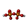
| #9: Chemical | ChemComp-ATP / #10: Chemical | #11: Chemical | ChemComp-ADP / | #12: Chemical | ChemComp-POP / | #13: Chemical | #14: Water | ChemComp-HOH / | |
|---|
-Details
| Has ligand of interest | N |
|---|---|
| Has protein modification | N |
-Experimental details
-Experiment
| Experiment | Method: ELECTRON MICROSCOPY |
|---|---|
| EM experiment | Aggregation state: PARTICLE / 3D reconstruction method: single particle reconstruction |
- Sample preparation
| Component | Name: EsNdhsUC1+ATP / Type: COMPLEX Details: E = RNAP sN = Sigma N dhsU = dhsU promoter DNA C1 = NtrC1 Entity ID: #1-#8 / Source: MULTIPLE SOURCES |
|---|---|
| Molecular weight | Value: 0.63 MDa / Experimental value: NO |
| Source (natural) | Organism:  |
| Source (recombinant) | Organism:  |
| Buffer solution | pH: 8 Details: 40 mM Tris-HCl, pH 8/RT, 200 mM KCl, 10 mM MgCl2, 1 mM DTT; fluorinated fos-choline-8 (FC8F) added to a final concentration of 1.5 mM during grid preparation |
| Specimen | Embedding applied: NO / Shadowing applied: NO / Staining applied: NO / Vitrification applied: YES |
| Specimen support | Grid material: GOLD / Grid mesh size: 400 divisions/in. / Grid type: C-flat-1.2/1.3 |
| Vitrification | Instrument: FEI VITROBOT MARK IV / Cryogen name: ETHANE / Humidity: 100 % / Chamber temperature: 310 K |
- Electron microscopy imaging
| Experimental equipment |  Model: Titan Krios / Image courtesy: FEI Company |
|---|---|
| Microscopy | Model: TFS KRIOS |
| Electron gun | Electron source:  FIELD EMISSION GUN / Accelerating voltage: 300 kV / Illumination mode: FLOOD BEAM FIELD EMISSION GUN / Accelerating voltage: 300 kV / Illumination mode: FLOOD BEAM |
| Electron lens | Mode: BRIGHT FIELD / Nominal defocus max: 2200 nm / Nominal defocus min: 800 nm |
| Specimen holder | Cryogen: NITROGEN |
| Image recording | Average exposure time: 1.4 sec. / Electron dose: 42 e/Å2 / Film or detector model: GATAN K3 BIOQUANTUM (6k x 4k) / Num. of grids imaged: 1 / Num. of real images: 17199 |
| Image scans | Width: 11520 / Height: 8184 |
- Processing
| EM software | Name: PHENIX / Version: 1.21.1_5286: / Category: model refinement |
|---|---|
| CTF correction | Type: PHASE FLIPPING AND AMPLITUDE CORRECTION |
| 3D reconstruction | Resolution: 2.7 Å / Resolution method: FSC 0.143 CUT-OFF / Num. of particles: 95920 / Symmetry type: POINT |
 Movie
Movie Controller


 PDBj
PDBj


