Journal: J Exp Med / Year: 2025 Title: Common cold embecovirus imprinting primes broadly neutralizing antibody responses to SARS-CoV-2 S2. Authors: Siriruk Changrob / Atsuhiro Yasuhara / Suncheol Park / Sandhya Bangaru / Lei Li / Chloe A Troxell / Peter J Halfmann / Steven A Erickson / Nicholas J Catanzaro / Meng Yuan / Panpan Zhou / ...Authors: Siriruk Changrob / Atsuhiro Yasuhara / Suncheol Park / Sandhya Bangaru / Lei Li / Chloe A Troxell / Peter J Halfmann / Steven A Erickson / Nicholas J Catanzaro / Meng Yuan / Panpan Zhou / Min Huang / G Dewey Wilbanks / Joshua J C McGrath / Gagandeep Singh / Sean A Nelson / Yanbin Fu / Nai-Ying Zheng / Sofia M Carayannopoulos / Haley L Dugan / Dustin G Shaw / Christopher T Stamper / Maria Lucia L Madariaga / Florian Krammer / Raiees Andrabi / Dennis R Burton / Andrew B Ward / Ian A Wilson / Yoshihiro Kawaoka / Patrick C Wilson / Abstract: The S2 subunit of the severe acute respiratory syndrome coronavirus 2 (SARS-CoV-2) spike is highly conserved across coronavirus strains and therefore is a potential pan-coronavirus vaccine target. ...The S2 subunit of the severe acute respiratory syndrome coronavirus 2 (SARS-CoV-2) spike is highly conserved across coronavirus strains and therefore is a potential pan-coronavirus vaccine target. However, antibodies targeting this region are typically non-neutralizing. We report herein that S2-targeting antibodies from patients who recovered from SARS-CoV-2 infection bound only closely related sarbecovirus subgenus strains and, like most known S2 antibodies, none of these were neutralizing. In contrast, first-exposure, severe acutely infected COVID-19 patients predominantly induced back-boosted antibody-secreting cells imprinted against past common cold coronavirus strain OC43 that were cross-reactive to as many as five subgenera of betacoronavirus strains and gave rise to antibodies that were neutralizing and protective. The antibodies targeted two different sites: one defined by competition with stem helix antibodies, and the second to an underdescribed epitope at the apex of S2. These findings suggest that S2-targeted vaccines could strategically exploit controlled OC43 priming followed by SARS-CoV-2 boosting to enhance the breadth and quality of protective antibody responses.
In the structure databanks used in Yorodumi, some data are registered as the other names, "COVID-19 virus" and "2019-nCoV". Here are the details of the virus and the list of structure data.
Jan 31, 2019. EMDB accession codes are about to change! (news from PDBe EMDB page)
EMDB accession codes are about to change! (news from PDBe EMDB page)
The allocation of 4 digits for EMDB accession codes will soon come to an end. Whilst these codes will remain in use, new EMDB accession codes will include an additional digit and will expand incrementally as the available range of codes is exhausted. The current 4-digit format prefixed with “EMD-” (i.e. EMD-XXXX) will advance to a 5-digit format (i.e. EMD-XXXXX), and so on. It is currently estimated that the 4-digit codes will be depleted around Spring 2019, at which point the 5-digit format will come into force.
The EM Navigator/Yorodumi systems omit the EMD- prefix.
Related info.:Q: What is EMD? / ID/Accession-code notation in Yorodumi/EM Navigator
Yorodumi is a browser for structure data from EMDB, PDB, SASBDB, etc.
This page is also the successor to EM Navigator detail page, and also detail information page/front-end page for Omokage search.
The word "yorodu" (or yorozu) is an old Japanese word meaning "ten thousand". "mi" (miru) is to see.
Related info.:EMDB / PDB / SASBDB / Comparison of 3 databanks / Yorodumi Search / Aug 31, 2016. New EM Navigator & Yorodumi / Yorodumi Papers / Jmol/JSmol / Function and homology information / Changes in new EM Navigator and Yorodumi
 Movie
Movie Controller


 Open data
 Basic information
 Map data
 Sample
 Keywords
 Function and homology information
 Homo sapiens (human) /
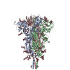
 Severe acute respiratory syndrome coronavirus 2
 Authors
 United States, 1 items
  Citation

 Structure visualization
 Downloads & links
 emd_48550.png
 http://ftp.pdbj.org/pub/emdb/structures/EMD-48550


 Z (Sec.)
 Y (Row.)
 X (Col.)


 Sample components
 Processing
 Electron microscopy
 FIELD EMISSION GUN