 Movie
Movie Controller

[English] 日本語
 Yorodumi
- EMDB-48544: Structure of mycobacterial NDH2 (type II NADH:quinone oxidoreductase) -
+ Open data

- Basic information
| Entry |  | |||||||||
|---|---|---|---|---|---|---|---|---|---|---|
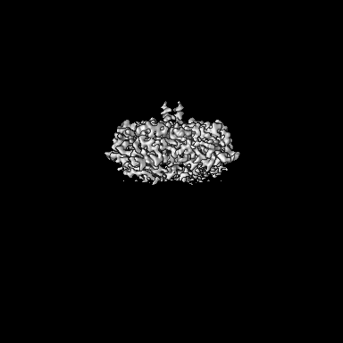
| Title | Structure of mycobacterial NDH2 (type II NADH:quinone oxidoreductase) | |||||||||
 Map data Map data | ||||||||||
 Sample Sample |
| |||||||||
 Keywords Keywords | flavoprotein / oxidoreductase / electron transport / metabolism / MEMBRANE PROTEIN | |||||||||
| Function / homology | Alternative NADH dehydrogenase / NADH:quinone reductase (non-electrogenic) / NADH dehydrogenase (quinone) (non-electrogenic) activity / FAD/NAD(P)-binding domain / Pyridine nucleotide-disulphide oxidoreductase / FAD/NAD(P)-binding domain superfamily / membrane / NADH:ubiquinone reductase (non-electrogenic) Function and homology information Function and homology information | |||||||||
| Biological species |  Mycolicibacterium smegmatis MC2 155 (bacteria) Mycolicibacterium smegmatis MC2 155 (bacteria) | |||||||||
| Method | single particle reconstruction / cryo EM / Resolution: 3.0 Å | |||||||||
 Authors Authors | Liang Y / Rubinstein JL | |||||||||
| Funding support |  Canada, 1 items Canada, 1 items
| |||||||||
 Citation Citation | Journal: J Med Chem / Year: 2025 Title: Structure of Mycobacterial NDH-2 Bound to a 2-Mercapto-Quinazolinone Inhibitor. Authors: Yingke Liang / Stephanie A Bueler / Gregory M Cook / John L Rubinstein /   Abstract: Mycobacterial type II NADH dehydrogenase (NDH-2) is a promising drug target because of its central role in energy metabolism in and other pathogens, and because it lacks a known mammalian homologue. ...Mycobacterial type II NADH dehydrogenase (NDH-2) is a promising drug target because of its central role in energy metabolism in and other pathogens, and because it lacks a known mammalian homologue. To facilitate optimization of lead compounds, we used electron cryomicroscopy (cryo-EM) to determine the structure of NDH-2 from , a fast-growing nonpathogenic model for respiration in . The structure shows that active mycobacterial NDH-2 is dimeric, with an arrangement of monomers in the dimer that differs from the arrangement described for other prokaryotic NDH-2 dimers, instead resembling dimers formed by NDH-2 in the eukaryotes and . A structure of the enzyme bound to a 2-mercapto-quinazolinone inhibitor shows that the compound interacts directly with the flavin adenine dinucleotide cofactor, blocking the menaquinone-reducing site. These results reveal structural elements of NDH-2 that could be used to design specific inhibitors of the mycobacterial enzyme. | |||||||||
| History |
|
- Structure visualization
| Supplemental images |
|---|

- Downloads & links
-EMDB archive
| Map data | emd_48544.map.gz | 107.3 MB | EMDB map data format | |
|---|---|---|---|---|
| Header (meta data) | emd-48544-v30.xmlemd-48544.xml | 19.6 KB 19.6 KB | Display Display | EMDB header |
| FSC (resolution estimation) | emd_48544_fsc.xml | 12.7 KB | Display | FSC data file |
| Images |  emd_48544.png emd_48544.png | 48.2 KB | ||
| Filedesc metadata | emd-48544.cif.gz | 6.3 KB | ||
| Others | emd_48544_half_map_1.map.gzemd_48544_half_map_2.map.gz | 200.3 MB 200.3 MB | ||
| Archive directory |  http://ftp.pdbj.org/pub/emdb/structures/EMD-48544ftp://ftp.pdbj.org/pub/emdb/structures/EMD-48544 http://ftp.pdbj.org/pub/emdb/structures/EMD-48544ftp://ftp.pdbj.org/pub/emdb/structures/EMD-48544 | HTTPS FTP |
-Validation report
| Summary document | emd_48544_validation.pdf.gz | 1.2 MB | Display | EMDB validaton report |
|---|---|---|---|---|
| Full document | emd_48544_full_validation.pdf.gz | 1.2 MB | Display | |
| Data in XML | emd_48544_validation.xml.gz | 21.6 KB | Display | |
| Data in CIF | emd_48544_validation.cif.gz | 27.8 KB | Display | |
| Arichive directory | https://ftp.pdbj.org/pub/emdb/validation_reports/EMD-48544ftp://ftp.pdbj.org/pub/emdb/validation_reports/EMD-48544 | HTTPS FTP |
-Related structure data
| Related structure data |  9mqyMC  9mqzC M: atomic model generated by this map C: citing same article ( |
|---|---|
| Similar structure data |

-Links
| EMDB pages | EMDB (EBI/PDBe) / EMDataResource |
|---|---|
| Related items in Molecule of the Month |


-Map
| File | Download / File: emd_48544.map.gz / Format: CCP4 / Size: 216 MB / Type: IMAGE STORED AS FLOATING POINT NUMBER (4 BYTES) | ||||||||||||||||||||||||||||||||||||
|---|---|---|---|---|---|---|---|---|---|---|---|---|---|---|---|---|---|---|---|---|---|---|---|---|---|---|---|---|---|---|---|---|---|---|---|---|---|
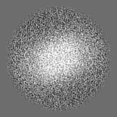
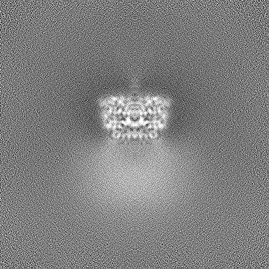
| Projections & slices | Image control
Images are generated by Spider. | ||||||||||||||||||||||||||||||||||||
| Voxel size | X=Y=Z: 0.64 Å | ||||||||||||||||||||||||||||||||||||
| Density |
| ||||||||||||||||||||||||||||||||||||
| Symmetry | Space group: 1 | ||||||||||||||||||||||||||||||||||||
| Details | EMDB XML:
|
 Z (Sec.)
 Y (Row.)
 X (Col.)


-Supplemental data
-Half map: #2
| File | emd_48544_half_map_1.map | ||||||||||||
|---|---|---|---|---|---|---|---|---|---|---|---|---|---|
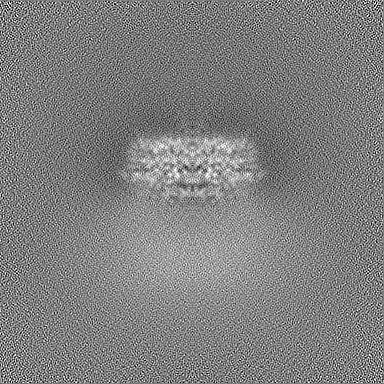
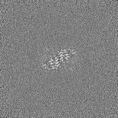
| Projections & Slices |
| ||||||||||||
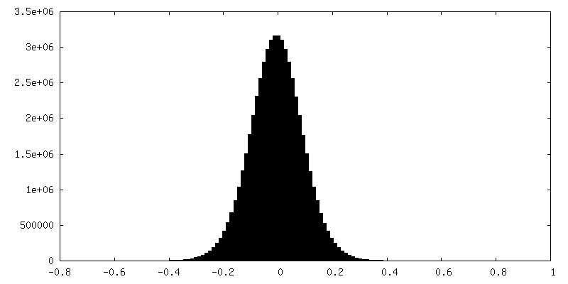
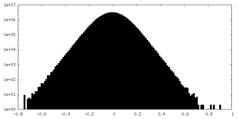
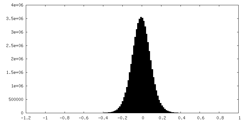
| Density Histograms |


-Half map: #1
| File | emd_48544_half_map_2.map | ||||||||||||
|---|---|---|---|---|---|---|---|---|---|---|---|---|---|
| Projections & Slices |
| ||||||||||||
| Density Histograms |


- Sample components
-Entire : NDH2 (non-electrogenic NADH:quinone oxidoreductase) from Mycobact...
| Entire | Name: NDH2 (non-electrogenic NADH:quinone oxidoreductase) from Mycobacterium smegmatis |
|---|---|
| Components |
|
-Supramolecule #1: NDH2 (non-electrogenic NADH:quinone oxidoreductase) from Mycobact...
| Supramolecule | Name: NDH2 (non-electrogenic NADH:quinone oxidoreductase) from Mycobacterium smegmatis type: complex / ID: 1 / Parent: 0 / Macromolecule list: #1 Details: Homodimer composed of two monomers of NDH2 from Mycobacterium smegmatis. |
|---|---|
| Source (natural) | Organism: Mycolicibacterium smegmatis MC2 155 (bacteria) / Synthetically produced: Yes |
| Molecular weight | Theoretical: 49 KDa |
-Macromolecule #1: NADH:ubiquinone reductase (non-electrogenic)
| Macromolecule | Name: NADH:ubiquinone reductase (non-electrogenic) / type: protein_or_peptide / ID: 1 Details: Includes sequence for a linker followed by a 3xFLAG affinity tag at the C terminus,Includes sequence for a linker followed by a 3xFLAG affinity tag at the C terminus Number of copies: 2 / Enantiomer: LEVO / EC number: NADH:quinone reductase (non-electrogenic) |
|---|---|
| Source (natural) | Organism: Mycolicibacterium smegmatis MC2 155 (bacteria) |
| Molecular weight | Theoretical: 53.14532 KDa |
| Sequence | String: MSHPGATASD RHKVVIIGSG FGGLTAAKTL KRADVDVKLI ARTTHHLFQP LLYQVATGII SEGEIAPATR VILRKQKNAQ VLLGDVTHI DLENKTVDSV LLGHTYSTPY DSLIIAAGAG QSYFGNDHFA EFAPGMKSID DALELRGRIL GAFEQAERSS D PVRRAKLL ...String: MSHPGATASD RHKVVIIGSG FGGLTAAKTL KRADVDVKLI ARTTHHLFQP LLYQVATGII SEGEIAPATR VILRKQKNAQ VLLGDVTHI DLENKTVDSV LLGHTYSTPY DSLIIAAGAG QSYFGNDHFA EFAPGMKSID DALELRGRIL GAFEQAERSS D PVRRAKLL TFTVVGAGPT GVEMAGQIAE LADQTLRGSF RHIDPTEARV ILLDAAPAVL PPMGEKLGKK ARARLEKMGV EV QLGAMVT DVDRNGITVK DSDGTIRRIE SACKVWSAGV SASPLGKDLA EQSGVELDRA GRVKVQPDLT LPGHPNVFVV GDM AAVEGV PGVAQGAIQG GRYAAKIIKR EVSGTSPKIR TPFEYFDKGS MATVSRFSAV AKVGPVEFAG FFAWLCWLVL HLVY LVGFK TKIVTLLSWG VTFLSTKRGQ LTITEQQAYA RTRIEELEEI AAAVQDTEKA ASGLSGQPPR SPSSGSSDYK DHDGD YKDH DIDYKDDDDK UniProtKB: NADH:ubiquinone reductase (non-electrogenic) |
-Macromolecule #2: FLAVIN-ADENINE DINUCLEOTIDE
| Macromolecule | Name: FLAVIN-ADENINE DINUCLEOTIDE / type: ligand / ID: 2 / Number of copies: 2 / Formula: FAD |
|---|---|
| Molecular weight | Theoretical: 785.55 Da |
| Chemical component information |  ChemComp-FAD: |
-Experimental details
-Structure determination
| Method | cryo EM |
|---|---|
 Processing Processing | single particle reconstruction |
| Aggregation state | particle |
-Sample preparation
| Buffer | pH: 6.8 |
|---|---|
| Grid | Model: Homemade / Material: COPPER/RHODIUM / Mesh: 400 / Support film - Material: GOLD / Support film - topology: HOLEY / Support film - Film thickness: 35 / Pretreatment - Type: GLOW DISCHARGE / Pretreatment - Time: 15 sec. |
| Vitrification | Cryogen name: ETHANE / Chamber humidity: 95 % / Chamber temperature: 277 K / Instrument: LEICA EM GP |
- Electron microscopy
| Microscope | TFS KRIOS |
|---|---|
| Image recording | Film or detector model: TFS FALCON 4i (4k x 4k) / Average electron dose: 70.0 e/Å2 |
| Electron beam | Acceleration voltage: 300 kV / Electron source:  FIELD EMISSION GUN FIELD EMISSION GUN |
| Electron optics | Illumination mode: FLOOD BEAM / Imaging mode: BRIGHT FIELD / Nominal defocus max: 2.0 µm / Nominal defocus min: 1.0 µm / Nominal magnification: 120000 |
| Experimental equipment |  Model: Titan Krios / Image courtesy: FEI Company |

