National Institutes of Health/National Cancer Institute (NIH/NCI)
CA92584
United States
Citation
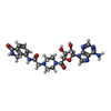
Journal: Nat Commun / Year: 2026 Title: PARP1-HPF1 structure and dynamics on nicked DNA suggest a mechanism for acute and localized ADP-ribosylation. Authors: Aleksandr Sverzhinsky / Huijun Xue / Marie-France Langelier / Marcelo V Muniz Corrêa / Joshua Del Mundo / Scott Classen / Michal Hammel / Eli Rothenberg / John M Pascal / Abstract: PARP1 detection of DNA strand breaks allosterically leads to PARP1 synthesis of poly(ADP-ribose) modifications that signal DNA damage. HPF1 engages activated PARP1 to control modification site ...PARP1 detection of DNA strand breaks allosterically leads to PARP1 synthesis of poly(ADP-ribose) modifications that signal DNA damage. HPF1 engages activated PARP1 to control modification site selection. Understanding of the mechanism of DNA break detection and catalytic activation is incomplete, due largely to limited structural information for full-length PARP1. Here, single-particle cryo-EM provides views of the full complement of PARP1 domains engaging a DNA single-strand break in the presence of HPF1 and a fragment of binding partner Timeless. Cryo-EM, single-molecule DNA dynamics, and small-angle X-ray scattering analysis indicate that PARP1 remains dynamic even when the multi-domain structure is organized on a DNA break, with the minimal catalytic region displaying high mobility relative to domains engaging damage. We propose that the organization of PARP1 domains on a DNA break releases a tethered, constitutively active catalytic region to modify molecules in a radius surrounding the DNA break site.
Supramolecule #1: Full-length human PARP1 bound to nicked DNA and in complex with H...
Supramolecule
Name: Full-length human PARP1 bound to nicked DNA and in complex with HPF1 and Timeless fragment type: complex / ID: 1 / Parent: 0 / Macromolecule list: #1-#2
In the structure databanks used in Yorodumi, some data are registered as the other names, "COVID-19 virus" and "2019-nCoV". Here are the details of the virus and the list of structure data.
Jan 31, 2019. EMDB accession codes are about to change! (news from PDBe EMDB page)
EMDB accession codes are about to change! (news from PDBe EMDB page)
The allocation of 4 digits for EMDB accession codes will soon come to an end. Whilst these codes will remain in use, new EMDB accession codes will include an additional digit and will expand incrementally as the available range of codes is exhausted. The current 4-digit format prefixed with “EMD-” (i.e. EMD-XXXX) will advance to a 5-digit format (i.e. EMD-XXXXX), and so on. It is currently estimated that the 4-digit codes will be depleted around Spring 2019, at which point the 5-digit format will come into force.
The EM Navigator/Yorodumi systems omit the EMD- prefix.
Related info.:Q: What is EMD? / ID/Accession-code notation in Yorodumi/EM Navigator
Yorodumi is a browser for structure data from EMDB, PDB, SASBDB, etc.
This page is also the successor to EM Navigator detail page, and also detail information page/front-end page for Omokage search.
The word "yorodu" (or yorozu) is an old Japanese word meaning "ten thousand". "mi" (miru) is to see.
Related info.:EMDB / PDB / SASBDB / Comparison of 3 databanks / Yorodumi Search / Aug 31, 2016. New EM Navigator & Yorodumi / Yorodumi Papers / Jmol/JSmol / Function and homology information / Changes in new EM Navigator and Yorodumi
 Movie
Movie Controller


 Open data
 Basic information
 Map data
 Sample
 Keywords
 Function and homology information
 Homo sapiens (human)
 Authors
 Canada,
  United States, 2 items
  Citation
 Structure visualization
 Downloads & links
 emd_48313.png
 http://ftp.pdbj.org/pub/emdb/structures/EMD-48313


 Z (Sec.)
 Y (Row.)
 X (Col.)


 Sample components
 Processing
 Electron microscopy
 FIELD EMISSION GUN
