 Movie
Movie Controller


+ Open data

- Basic information
| Entry |  | |||||||||
|---|---|---|---|---|---|---|---|---|---|---|
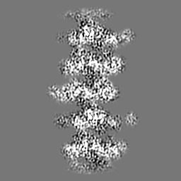
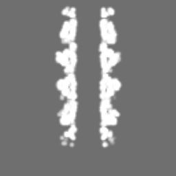
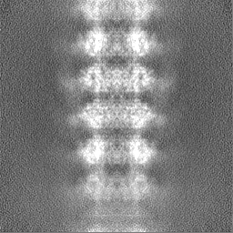
| Title | cryoEM structure of Escherichia phage YDC107 tail core | |||||||||
 Map data Map data | ||||||||||
 Sample Sample |
| |||||||||
 Keywords Keywords | phage tail / bacteriophage / E. coli phage / YDC107 / helical tail / VIRUS / VIRAL PROTEIN | |||||||||
| Function / homology | Lambda phage tail tube protein / Lambda phage tail tube protein, TTP / Bacterial Ig-like domain (group 2) / Invasin/intimin cell-adhesion fragments / Bacterial Ig-like domain 2 / Bacterial Ig-like domain, group 2 / Major tail protein V Function and homology information Function and homology information | |||||||||
| Biological species |  Escherichia phage YDC107_1 (virus) Escherichia phage YDC107_1 (virus) | |||||||||
| Method | helical reconstruction / cryo EM / Resolution: 3.22 Å | |||||||||
 Authors Authors | Kopylov M | |||||||||
| Funding support |  United States, 1 items United States, 1 items
| |||||||||
 Citation Citation | Journal: To Be Published Title: Identification and cryoEM structure determination of Escherichia phage YDC107 tail found in a bacteria-contaminated buffer Authors: Kopylov M / Jenkins MC | |||||||||
| History |
|
- Structure visualization
| Supplemental images |
|---|

- Downloads & links
-EMDB archive
| Map data | emd_48226.map.gz | 19.5 MB | EMDB map data format | |
|---|---|---|---|---|
| Header (meta data) | emd-48226-v30.xmlemd-48226.xml | 15.2 KB 15.2 KB | Display Display | EMDB header |
| FSC (resolution estimation) | emd_48226_fsc.xml | 8.4 KB | Display | FSC data file |
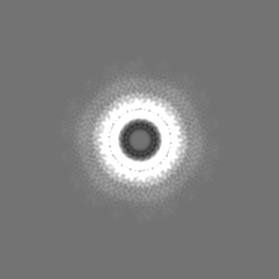
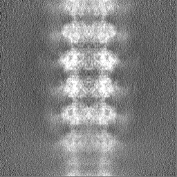
| Images |  emd_48226.png emd_48226.png | 111.3 KB | ||
| Masks | emd_48226_msk_1.map | 64 MB | Mask map | |
| Filedesc metadata | emd-48226.cif.gz | 5.6 KB | ||
| Others | emd_48226_half_map_1.map.gzemd_48226_half_map_2.map.gz | 59.3 MB 59.3 MB | ||
| Archive directory |  http://ftp.pdbj.org/pub/emdb/structures/EMD-48226ftp://ftp.pdbj.org/pub/emdb/structures/EMD-48226 http://ftp.pdbj.org/pub/emdb/structures/EMD-48226ftp://ftp.pdbj.org/pub/emdb/structures/EMD-48226 | HTTPS FTP |
-Related structure data
| Related structure data |  9mfeMC M: atomic model generated by this map C: citing same article ( |
|---|---|
| Similar structure data |
-Links
| EMDB pages | EMDB (EBI/PDBe) / EMDataResource |
|---|
-Map
| File | Download / File: emd_48226.map.gz / Format: CCP4 / Size: 64 MB / Type: IMAGE STORED AS FLOATING POINT NUMBER (4 BYTES) | ||||||||||||||||||||||||||||||||||||
|---|---|---|---|---|---|---|---|---|---|---|---|---|---|---|---|---|---|---|---|---|---|---|---|---|---|---|---|---|---|---|---|---|---|---|---|---|---|
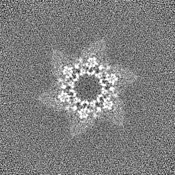
| Projections & slices | Image control
Images are generated by Spider. | ||||||||||||||||||||||||||||||||||||
| Voxel size | X=Y=Z: 1.058 Å | ||||||||||||||||||||||||||||||||||||
| Density |
| ||||||||||||||||||||||||||||||||||||
| Symmetry | Space group: 1 | ||||||||||||||||||||||||||||||||||||
| Details | EMDB XML:
|
 Z (Sec.)
 Y (Row.)
 X (Col.)


-Supplemental data
-Mask #1
| File | emd_48226_msk_1.map | ||||||||||||
|---|---|---|---|---|---|---|---|---|---|---|---|---|---|
| Projections & Slices |
| ||||||||||||
| Density Histograms |


-Half map: #2
| File | emd_48226_half_map_1.map | ||||||||||||
|---|---|---|---|---|---|---|---|---|---|---|---|---|---|
| Projections & Slices |
| ||||||||||||
| Density Histograms |


-Half map: #1
| File | emd_48226_half_map_2.map | ||||||||||||
|---|---|---|---|---|---|---|---|---|---|---|---|---|---|
| Projections & Slices |
| ||||||||||||
| Density Histograms |


- Sample components
-Entire : Escherichia phage YDC107_1
| Entire | Name: Escherichia phage YDC107_1 (virus) |
|---|---|
| Components |
|
-Supramolecule #1: Escherichia phage YDC107_1
| Supramolecule | Name: Escherichia phage YDC107_1 / type: complex / ID: 1 / Parent: 0 / Macromolecule list: all Details: Tail fragments were identified in the contaminated buffer during cryoEM sample screening |
|---|---|
| Source (natural) | Organism: Escherichia phage YDC107_1 (virus) |
-Macromolecule #1: Major tail protein V
| Macromolecule | Name: Major tail protein V / type: protein_or_peptide / ID: 1 Details: Residues 155 and onwards are not resolved to high resolution and are excluded from the model Number of copies: 6 / Enantiomer: LEVO |
|---|---|
| Source (natural) | Organism: Escherichia phage YDC107_1 (virus) |
| Molecular weight | Theoretical: 25.699557 KDa |
| Sequence | String: MPTPNPLPVK GAGTTLWVYK GNGDPYANPL SDVDWSRLAK VKDLTPGELT AESYDDSYLD DEDADWTATG QGQKSAGDTS FTLAWMPGE QGQQALLAWF NEGDTRAYKI RFPNGTVDVF RGWVSSIGKA VTAKEVITRT VKVTNVGRPS MAEDRSTVTA A TGMTVTPA ...String: MPTPNPLPVK GAGTTLWVYK GNGDPYANPL SDVDWSRLAK VKDLTPGELT AESYDDSYLD DEDADWTATG QGQKSAGDTS FTLAWMPGE QGQQALLAWF NEGDTRAYKI RFPNGTVDVF RGWVSSIGKA VTAKEVITRT VKVTNVGRPS MAEDRSTVTA A TGMTVTPA SASVVKGQST TLTVAFQPEG ATDKSFRAVS SDKTKATVSV SGMTITVNGV AAGKVNIPVV SGNGEFAAVA EI TVTAS UniProtKB: Major tail protein V |
-Experimental details
-Structure determination
| Method | cryo EM |
|---|---|
 Processing Processing | helical reconstruction |
| Aggregation state | filament |
-Sample preparation
| Buffer | pH: 7 |
|---|---|
| Grid | Model: Quantifoil R1.2/1.3 / Material: COPPER / Mesh: 300 / Support film - Material: CARBON / Support film - topology: HOLEY / Pretreatment - Type: PLASMA CLEANING / Pretreatment - Time: 15 sec. / Pretreatment - Atmosphere: OTHER |
| Vitrification | Cryogen name: ETHANE / Chamber humidity: 95 % / Chamber temperature: 300 K / Instrument: FEI VITROBOT MARK IV / Details: blot force 1 blot time 6.5 s. |
- Electron microscopy
| Microscope | TFS KRIOS |
|---|---|
| Specialist optics | Energy filter - Name: GIF Bioquantum / Energy filter - Slit width: 20 eV |
| Image recording | Film or detector model: GATAN K3 BIOQUANTUM (6k x 4k) / Number grids imaged: 1 / Average electron dose: 54.0 e/Å2 |
| Electron beam | Acceleration voltage: 300 kV / Electron source:  FIELD EMISSION GUN FIELD EMISSION GUN |
| Electron optics | C2 aperture diameter: 100.0 µm / Illumination mode: FLOOD BEAM / Imaging mode: BRIGHT FIELD / Cs: 2.7 mm / Nominal defocus max: 2.0 µm / Nominal defocus min: 1.0 µm / Nominal magnification: 81000 |
| Sample stage | Specimen holder model: FEI TITAN KRIOS AUTOGRID HOLDER / Cooling holder cryogen: NITROGEN |
| Experimental equipment |  Model: Titan Krios / Image courtesy: FEI Company |
-Image processing
| Final reconstruction | Applied symmetry - Helical parameters - Δz: 41.77 Å Applied symmetry - Helical parameters - Δ&Phi: 17.90 ° Applied symmetry - Helical parameters - Axial symmetry: C6 (6 fold cyclic) Algorithm: FOURIER SPACE / Resolution.type: BY AUTHOR / Resolution: 3.22 Å / Resolution method: FSC 0.143 CUT-OFF / Number images used: 5927 |
|---|---|
| Startup model | Type of model: NONE |
| Final angle assignment | Type: NOT APPLICABLE / Software - Name: cryoSPARC |
| FSC plot (resolution estimation) |  |
-Atomic model buiding 1
| Initial model | Chain - Source name: Other / Chain - Initial model type: experimental model / Details: Modelangelo |
|---|---|
| Refinement | Space: REAL / Protocol: FLEXIBLE FIT |
| Output model | PDB-9mfe: |
