Movie
Movie Controller

[English] 日本語
 Yorodumi
- EMDB-48153: Cryo-EM structure of SARS-CoV-2 Omicron JN.1.11 spike protein (on... -
+ Open data

- Basic information
| Entry |  | |||||||||
|---|---|---|---|---|---|---|---|---|---|---|
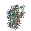
| Title | Cryo-EM structure of SARS-CoV-2 Omicron JN.1.11 spike protein (one RBD up state) | |||||||||
 Map data Map data | structure of SARS-CoV-2 Omicron JN.1.11 spike protein (one RBD up state) | |||||||||
 Sample Sample |
| |||||||||
 Keywords Keywords | SARS-CoV-2 / COVID-19 / Spike / VIRAL PROTEIN | |||||||||
| Function / homology |  Function and homology information Function and homology informationpositive regulation of viral entry into host cell / host cell endoplasmic reticulum-Golgi intermediate compartment membrane / receptor-mediated virion attachment to host cell / host cell surface receptor binding / endocytosis involved in viral entry into host cell / fusion of virus membrane with host plasma membrane / fusion of virus membrane with host endosome membrane / viral envelope / host cell plasma membrane / virion membrane ...positive regulation of viral entry into host cell / host cell endoplasmic reticulum-Golgi intermediate compartment membrane / receptor-mediated virion attachment to host cell / host cell surface receptor binding / endocytosis involved in viral entry into host cell / fusion of virus membrane with host plasma membrane / fusion of virus membrane with host endosome membrane / viral envelope / host cell plasma membrane / virion membrane / membrane / identical protein binding Similarity search - Function | |||||||||
| Biological species |   Severe acute respiratory syndrome coronavirus 2 Severe acute respiratory syndrome coronavirus 2 | |||||||||
| Method | single particle reconstruction / cryo EM / Resolution: 2.93 Å | |||||||||
 Authors Authors | Feng Z / Huang J / Ward AB | |||||||||
| Funding support |  United States, 1 items United States, 1 items
| |||||||||
 Citation Citation | Journal: Cell Rep / Year: 2025 Title: Structural and functional insights into the evolution of SARS-CoV-2 KP.3.1.1 spike protein. Authors: Ziqi Feng / Jiachen Huang / Sabyasachi Baboo / Jolene K Diedrich / Sandhya Bangaru / James C Paulson / John R Yates / Meng Yuan / Ian A Wilson / Andrew B Ward / Abstract: The JN.1-sublineage KP.3.1.1 recently emerged as the globally prevalent SARS-CoV-2 variant, demonstrating increased infectivity and antibody escape. We investigate how mutations and a deletion in the ...The JN.1-sublineage KP.3.1.1 recently emerged as the globally prevalent SARS-CoV-2 variant, demonstrating increased infectivity and antibody escape. We investigate how mutations and a deletion in the KP.3.1.1 spike protein (S) affect hACE2 binding and antibody escape. Mass spectrometry confirms a new glycan site at residue N30 that alters the glycoforms at neighboring N61. Cryoelectron microscopy (cryo-EM) structures show that the N30 glycan and rearrangement of adjacent residues do not significantly change the overall spike structure, up-down ratio of receptor-binding domains (RBDs), or hACE2 binding. Furthermore, a KP.3.1.1 S with hACE2 structure further confirms an epistatic effect between F456L and Q493E on hACE2 binding. Our analysis shows that SARS-CoV-2 variants that emerged after late 2023 are now incorporating reversions to residues found in other sarbecoviruses, including the N30 glycan, Q493E, and others. Overall, these results inform on the structural and functional consequences of the KP.3.1.1 mutations, the current SARS-CoV-2 evolutionary trajectory, and immune evasion. | |||||||||
| History |
|
- Structure visualization
| Supplemental images |
|---|

- Downloads & links
-EMDB archive
| Map data | emd_48153.map.gz | 484.1 MB | EMDB map data format | |
|---|---|---|---|---|
| Header (meta data) | emd-48153-v30.xmlemd-48153.xml | 19.3 KB 19.3 KB | Display Display | EMDB header |
| FSC (resolution estimation) | emd_48153_fsc.xml | 17 KB | Display | FSC data file |
| Images |  emd_48153.png emd_48153.png | 86.2 KB | ||
| Masks | emd_48153_msk_1.map | 512 MB | Mask map | |
| Filedesc metadata | emd-48153.cif.gz | 6.8 KB | ||
| Others | emd_48153_half_map_1.map.gzemd_48153_half_map_2.map.gz | 475.1 MB 475.1 MB | ||
| Archive directory |  http://ftp.pdbj.org/pub/emdb/structures/EMD-48153ftp://ftp.pdbj.org/pub/emdb/structures/EMD-48153 http://ftp.pdbj.org/pub/emdb/structures/EMD-48153ftp://ftp.pdbj.org/pub/emdb/structures/EMD-48153 | HTTPS FTP |
-Related structure data
| Related structure data |  9ellMC  9eleC  9elfC  9elgC  9elhC  9eliC  9eljC  9elkC  9elmC  9elnC  9eloC  9elpC  9elqC M: atomic model generated by this map C: citing same article ( |
|---|---|
| Similar structure data |


-Links
| EMDB pages | EMDB (EBI/PDBe) / EMDataResource |
|---|
-Map
| File | Download / File: emd_48153.map.gz / Format: CCP4 / Size: 512 MB / Type: IMAGE STORED AS FLOATING POINT NUMBER (4 BYTES) | ||||||||||||||||||||||||||||||||||||
|---|---|---|---|---|---|---|---|---|---|---|---|---|---|---|---|---|---|---|---|---|---|---|---|---|---|---|---|---|---|---|---|---|---|---|---|---|---|
| Annotation | structure of SARS-CoV-2 Omicron JN.1.11 spike protein (one RBD up state) | ||||||||||||||||||||||||||||||||||||
| Projections & slices | Image control
Images are generated by Spider. | ||||||||||||||||||||||||||||||||||||
| Voxel size | X=Y=Z: 0.718 Å | ||||||||||||||||||||||||||||||||||||
| Density |
| ||||||||||||||||||||||||||||||||||||
| Symmetry | Space group: 1 | ||||||||||||||||||||||||||||||||||||
| Details | EMDB XML:
|
 Z (Sec.)
 Y (Row.)
 X (Col.)


-Supplemental data
-Mask #1
| File | emd_48153_msk_1.map | ||||||||||||
|---|---|---|---|---|---|---|---|---|---|---|---|---|---|
| Projections & Slices |
| ||||||||||||
| Density Histograms |


-Half map: Half Map B
| File | emd_48153_half_map_1.map | ||||||||||||
|---|---|---|---|---|---|---|---|---|---|---|---|---|---|
| Annotation | Half Map B | ||||||||||||
| Projections & Slices |
| ||||||||||||
| Density Histograms |


-Half map: Half Map A
| File | emd_48153_half_map_2.map | ||||||||||||
|---|---|---|---|---|---|---|---|---|---|---|---|---|---|
| Annotation | Half Map A | ||||||||||||
| Projections & Slices |
| ||||||||||||
| Density Histograms |


- Sample components
-Entire : SARS-CoV-2 Omicron JN.1.11 spike protein
| Entire | Name: SARS-CoV-2 Omicron JN.1.11 spike protein |
|---|---|
| Components |
|
-Supramolecule #1: SARS-CoV-2 Omicron JN.1.11 spike protein
| Supramolecule | Name: SARS-CoV-2 Omicron JN.1.11 spike protein / type: complex / ID: 1 / Parent: 0 / Macromolecule list: #1 |
|---|---|
| Source (natural) | Organism: Severe acute respiratory syndrome coronavirus 2 |
-Macromolecule #1: Spike glycoprotein
| Macromolecule | Name: Spike glycoprotein / type: protein_or_peptide / ID: 1 / Number of copies: 3 / Enantiomer: LEVO |
|---|---|
| Source (natural) | Organism: Severe acute respiratory syndrome coronavirus 2 |
| Molecular weight | Theoretical: 133.414234 KDa |
| Recombinant expression | Organism:  Homo sapiens (human) Homo sapiens (human) |
| Sequence | String: MFVFLVLLPL VSSQCVNLIT TTQSYTNSFT RGVYYPDKVF RSSVLHLTQD LFLPFFSNVT WFHAISGTNG TKRFDNPVLP FNDGVYFAS TEKSNIIRGW IFGTTLDSKT QSLLIVNNAT NVFIKVCEFQ FCNDPFLDVY HKNNKSWMES ESGVYSSANN C TFEYVSQP ...String: MFVFLVLLPL VSSQCVNLIT TTQSYTNSFT RGVYYPDKVF RSSVLHLTQD LFLPFFSNVT WFHAISGTNG TKRFDNPVLP FNDGVYFAS TEKSNIIRGW IFGTTLDSKT QSLLIVNNAT NVFIKVCEFQ FCNDPFLDVY HKNNKSWMES ESGVYSSANN C TFEYVSQP FLMDLEGKQG NFKNLREFVF KNIDGYFKIY SKHTPINIGR DFPQGFSALE PLVDLPIGIN ITRFQTLLAL NR SYLTPGD SSSGWTAGAA DYYVGYLQPR TFLLKYNENG TITDAVDCAL DPLSETKCTL KSFTVEKGIY QTSNFRVQPT ESI VRFPNV TNLCPFHEVF NATRFASVYA WNRTRISNCV ADYSVLYNFA PFFAFKCYGV SPTKLNDLCF TNVYADSFVI KGNE VSQIA PGQTGNIADY NYKLPDDFTG CVIAWNSNKL DSKHSGNYDY WYRSFRKSKL KPFERDISTE IYQAGNKPCK GKGPN CYFP LQSYGFRPTY GVGHQPYRVV VLSFELLHAP ATVCGPKKST NLVKNKCVNF NFNGLTGTGV LTKSNKKFLP FQQFGR DIV DTTDAVRDPQ TLEILDITPC SFGGVSVITP GTNTSNQVAV LYQGVNCTEV SVAIHADQLT PTWRVYSTGS NVFQTRA GC LIGAEYVNNS YECDIPIGAG ICASYQTQTK SRGSASSVAS QSIIAYTMSL GAENSVAYSN NSIAIPTNFT ISVTTEIL P VSMTKTSVDC TMYICGDSTE CSNLLLQYGS FCTQLKRALT GIAVEQDKNT QEVFAQVKQI YKTPPIKYFG GFNFSQILP DPSKPSKRSP IEDLLFNKVT LADAGFIKQY GDCLGDIAAR DLICAQKFNG LTVLPPLLTD EMIAQYTSAL LAGTITSGWT FGAGPALQI PFPMQMAYRF NGIGVTQNVL YENQKLIANQ FNSAIGKIQD SLFSTPSALG KLQDVVNHNA QALNTLVKQL S SKFGAISS VLNDILSRLD PPEAEVQIDR LITGRLQSLQ TYVTQQLIRA AEIRASANLA ATKMSECVLG QSKRVDFCGK GY HLMSFPQ SAPHGVVFLH VTYVPAQEKN FTTAPAICHD GKAHFPREGV FVSNGTHWFL TQRNFYEPQI ITTDNTFVSG NCD VVIGIV NNTVYDPLQL ELDSFKEELD KYFKNHTSPD VDLGDISGIN ASVVNIQKEI DRLNEVAKNL NESLIDLQEL GKYE Q UniProtKB: Spike glycoprotein |
-Macromolecule #4: 2-acetamido-2-deoxy-beta-D-glucopyranose
| Macromolecule | Name: 2-acetamido-2-deoxy-beta-D-glucopyranose / type: ligand / ID: 4 / Number of copies: 22 / Formula: NAG |
|---|---|
| Molecular weight | Theoretical: 221.208 Da |
| Chemical component information |  ChemComp-NAG: |
-Experimental details
-Structure determination
| Method | cryo EM |
|---|---|
 Processing Processing | single particle reconstruction |
| Aggregation state | particle |
-Sample preparation
| Buffer | pH: 7.4 |
|---|---|
| Vitrification | Cryogen name: ETHANE |
- Electron microscopy
| Microscope | TFS GLACIOS |
|---|---|
| Image recording | Film or detector model: FEI FALCON IV (4k x 4k) / Average electron dose: 44.84 e/Å2 |
| Electron beam | Acceleration voltage: 200 kV / Electron source:  FIELD EMISSION GUN FIELD EMISSION GUN |
| Electron optics | Illumination mode: FLOOD BEAM / Imaging mode: BRIGHT FIELD / Nominal defocus max: 1.7 µm / Nominal defocus min: 0.7000000000000001 µm |