Movie
Movie Controller

[English] 日本語
 Yorodumi
- EMDB-48151: Cryo-EM structure of SARS-CoV-2 Omicron JN.1.11+Q493E+S31deletion... -
+ Open data

- Basic information
| Entry |  | |||||||||
|---|---|---|---|---|---|---|---|---|---|---|
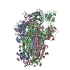
| Title | Cryo-EM structure of SARS-CoV-2 Omicron JN.1.11+Q493E+S31deletion spike protein (one RBD up state) | |||||||||
 Map data Map data | structure of SARS-CoV-2 Omicron JN.1.11+Q493E+S31deletion spike protein (one RBD up state) | |||||||||
 Sample Sample |
| |||||||||
 Keywords Keywords | SARS-CoV-2 / COVID-19 / Spike / VIRAL PROTEIN | |||||||||
| Function / homology |  Function and homology information Function and homology informationpositive regulation of viral entry into host cell / host cell endoplasmic reticulum-Golgi intermediate compartment membrane / receptor-mediated virion attachment to host cell / host cell surface receptor binding / endocytosis involved in viral entry into host cell / fusion of virus membrane with host plasma membrane / fusion of virus membrane with host endosome membrane / viral envelope / host cell plasma membrane / virion membrane ...positive regulation of viral entry into host cell / host cell endoplasmic reticulum-Golgi intermediate compartment membrane / receptor-mediated virion attachment to host cell / host cell surface receptor binding / endocytosis involved in viral entry into host cell / fusion of virus membrane with host plasma membrane / fusion of virus membrane with host endosome membrane / viral envelope / host cell plasma membrane / virion membrane / membrane / identical protein binding Similarity search - Function | |||||||||
| Biological species |   Severe acute respiratory syndrome coronavirus 2 Severe acute respiratory syndrome coronavirus 2 | |||||||||
| Method | single particle reconstruction / cryo EM / Resolution: 2.92 Å | |||||||||
 Authors Authors | Feng Z / Huang J / Ward AB | |||||||||
| Funding support |  United States, 1 items United States, 1 items
| |||||||||
 Citation Citation | Journal: Cell Rep / Year: 2025 Title: Structural and functional insights into the evolution of SARS-CoV-2 KP.3.1.1 spike protein. Authors: Ziqi Feng / Jiachen Huang / Sabyasachi Baboo / Jolene K Diedrich / Sandhya Bangaru / James C Paulson / John R Yates / Meng Yuan / Ian A Wilson / Andrew B Ward / Abstract: The JN.1-sublineage KP.3.1.1 recently emerged as the globally prevalent SARS-CoV-2 variant, demonstrating increased infectivity and antibody escape. We investigate how mutations and a deletion in the ...The JN.1-sublineage KP.3.1.1 recently emerged as the globally prevalent SARS-CoV-2 variant, demonstrating increased infectivity and antibody escape. We investigate how mutations and a deletion in the KP.3.1.1 spike protein (S) affect hACE2 binding and antibody escape. Mass spectrometry confirms a new glycan site at residue N30 that alters the glycoforms at neighboring N61. Cryoelectron microscopy (cryo-EM) structures show that the N30 glycan and rearrangement of adjacent residues do not significantly change the overall spike structure, up-down ratio of receptor-binding domains (RBDs), or hACE2 binding. Furthermore, a KP.3.1.1 S with hACE2 structure further confirms an epistatic effect between F456L and Q493E on hACE2 binding. Our analysis shows that SARS-CoV-2 variants that emerged after late 2023 are now incorporating reversions to residues found in other sarbecoviruses, including the N30 glycan, Q493E, and others. Overall, these results inform on the structural and functional consequences of the KP.3.1.1 mutations, the current SARS-CoV-2 evolutionary trajectory, and immune evasion. | |||||||||
| History |
|
- Structure visualization
| Supplemental images |
|---|

- Downloads & links
-EMDB archive
| Map data | emd_48151.map.gz | 484 MB | EMDB map data format | |
|---|---|---|---|---|
| Header (meta data) | emd-48151-v30.xmlemd-48151.xml | 19.4 KB 19.4 KB | Display Display | EMDB header |
| FSC (resolution estimation) | emd_48151_fsc.xml | 17 KB | Display | FSC data file |
| Images |  emd_48151.png emd_48151.png | 79.4 KB | ||
| Masks | emd_48151_msk_1.map | 512 MB | Mask map | |
| Filedesc metadata | emd-48151.cif.gz | 6.8 KB | ||
| Others | emd_48151_half_map_1.map.gzemd_48151_half_map_2.map.gz | 474.7 MB 474.7 MB | ||
| Archive directory |  http://ftp.pdbj.org/pub/emdb/structures/EMD-48151ftp://ftp.pdbj.org/pub/emdb/structures/EMD-48151 http://ftp.pdbj.org/pub/emdb/structures/EMD-48151ftp://ftp.pdbj.org/pub/emdb/structures/EMD-48151 | HTTPS FTP |
-Related structure data
| Related structure data |  9eljMC  9eleC  9elfC  9elgC  9elhC  9eliC  9elkC  9ellC  9elmC  9elnC  9eloC  9elpC  9elqC M: atomic model generated by this map C: citing same article ( |
|---|---|
| Similar structure data |


-Links
| EMDB pages | EMDB (EBI/PDBe) / EMDataResource |
|---|
-Map
| File | Download / File: emd_48151.map.gz / Format: CCP4 / Size: 512 MB / Type: IMAGE STORED AS FLOATING POINT NUMBER (4 BYTES) | ||||||||||||||||||||||||||||||||||||
|---|---|---|---|---|---|---|---|---|---|---|---|---|---|---|---|---|---|---|---|---|---|---|---|---|---|---|---|---|---|---|---|---|---|---|---|---|---|
| Annotation | structure of SARS-CoV-2 Omicron JN.1.11+Q493E+S31deletion spike protein (one RBD up state) | ||||||||||||||||||||||||||||||||||||
| Projections & slices | Image control
Images are generated by Spider. | ||||||||||||||||||||||||||||||||||||
| Voxel size | X=Y=Z: 0.718 Å | ||||||||||||||||||||||||||||||||||||
| Density |
| ||||||||||||||||||||||||||||||||||||
| Symmetry | Space group: 1 | ||||||||||||||||||||||||||||||||||||
| Details | EMDB XML:
|
 Z (Sec.)
 Y (Row.)
 X (Col.)


-Supplemental data
-Mask #1
| File | emd_48151_msk_1.map | ||||||||||||
|---|---|---|---|---|---|---|---|---|---|---|---|---|---|
| Projections & Slices |
| ||||||||||||
| Density Histograms |


-Half map: Half Map B
| File | emd_48151_half_map_1.map | ||||||||||||
|---|---|---|---|---|---|---|---|---|---|---|---|---|---|
| Annotation | Half Map B | ||||||||||||
| Projections & Slices |
| ||||||||||||
| Density Histograms |


-Half map: Half Map A
| File | emd_48151_half_map_2.map | ||||||||||||
|---|---|---|---|---|---|---|---|---|---|---|---|---|---|
| Annotation | Half Map A | ||||||||||||
| Projections & Slices |
| ||||||||||||
| Density Histograms |


- Sample components
-Entire : SARS-CoV-2 Omicron JN.1.11+Q493E+S31deletion spike protein
| Entire | Name: SARS-CoV-2 Omicron JN.1.11+Q493E+S31deletion spike protein |
|---|---|
| Components |
|
-Supramolecule #1: SARS-CoV-2 Omicron JN.1.11+Q493E+S31deletion spike protein
| Supramolecule | Name: SARS-CoV-2 Omicron JN.1.11+Q493E+S31deletion spike protein type: complex / ID: 1 / Parent: 0 / Macromolecule list: #1 |
|---|---|
| Source (natural) | Organism: Severe acute respiratory syndrome coronavirus 2 |
-Macromolecule #1: Spike glycoprotein
| Macromolecule | Name: Spike glycoprotein / type: protein_or_peptide / ID: 1 / Number of copies: 3 / Enantiomer: LEVO |
|---|---|
| Source (natural) | Organism: Severe acute respiratory syndrome coronavirus 2 |
| Molecular weight | Theoretical: 133.214031 KDa |
| Recombinant expression | Organism:  Homo sapiens (human) Homo sapiens (human) |
| Sequence | String: MFVFLVLLPL VSSQCVNLIT TTQSYTNFTR GVYYPDKVFR SSVLHLTQDL FLPFFSNVTW FHAISGTNGT KRFDNPVLPF NDGVYFAST EKSNIIRGWI FGTTLDSKTQ SLLIVNNATN VFIKVCEFQF CNDPFLDVYH KNNKSWMESE SGVYSSANNC T FEYVSQPF ...String: MFVFLVLLPL VSSQCVNLIT TTQSYTNFTR GVYYPDKVFR SSVLHLTQDL FLPFFSNVTW FHAISGTNGT KRFDNPVLPF NDGVYFAST EKSNIIRGWI FGTTLDSKTQ SLLIVNNATN VFIKVCEFQF CNDPFLDVYH KNNKSWMESE SGVYSSANNC T FEYVSQPF LMDLEGKQGN FKNLREFVFK NIDGYFKIYS KHTPIIGRDF PQGFSALEPL VDLPIGINIT RFQTLLALNR SY LTPGDSS SGWTAGAADY YVGYLQPRTF LLKYNENGTI TDAVDCALDP LSETKCTLKS FTVEKGIYQT SNFRVQPTES IVR FPNVTN LCPFHEVFNA TRFASVYAWN RTRISNCVAD YSVLYNFAPF FAFKCYGVSP TKLNDLCFTN VYADSFVIKG NEVS QIAPG QTGNIADYNY KLPDDFTGCV IAWNSNKLDS KHSGNYDYWY RSFRKSKLKP FERDISTEIY QAGNKPCKGK GPNCY FPLE SYGFRPTYGV GHQPYRVVVL SFELLHAPAT VCGPKKSTNL VKNKCVNFNF NGLTGTGVLT KSNKKFLPFQ QFGRDI VDT TDAVRDPQTL EILDITPCSF GGVSVITPGT NTSNQVAVLY QGVNCTEVSV AIHADQLTPT WRVYSTGSNV FQTRAGC LI GAEYVNNSYE CDIPIGAGIC ASYQTQTKSR GSASSVASQS IIAYTMSLGA ENSVAYSNNS IAIPTNFTIS VTTEILPV S MTKTSVDCTM YICGDSTECS NLLLQYGSFC TQLKRALTGI AVEQDKNTQE VFAQVKQIYK TPPIKYFGGF NFSQILPDP SKPSKRSPIE DLLFNKVTLA DAGFIKQYGD CLGDIAARDL ICAQKFNGLT VLPPLLTDEM IAQYTSALLA GTITSGWTFG AGPALQIPF PMQMAYRFNG IGVTQNVLYE NQKLIANQFN SAIGKIQDSL FSTPSALGKL QDVVNHNAQA LNTLVKQLSS K FGAISSVL NDILSRLDPP EAEVQIDRLI TGRLQSLQTY VTQQLIRAAE IRASANLAAT KMSECVLGQS KRVDFCGKGY HL MSFPQSA PHGVVFLHVT YVPAQEKNFT TAPAICHDGK AHFPREGVFV SNGTHWFLTQ RNFYEPQIIT TDNTFVSGNC DVV IGIVNN TVYDPLQLEL DSFKEELDKY FKNHTSPDVD LGDISGINAS VVNIQKEIDR LNEVAKNLNE SLIDLQELGK YEQ UniProtKB: Spike glycoprotein |
-Macromolecule #4: 2-acetamido-2-deoxy-beta-D-glucopyranose
| Macromolecule | Name: 2-acetamido-2-deoxy-beta-D-glucopyranose / type: ligand / ID: 4 / Number of copies: 22 / Formula: NAG |
|---|---|
| Molecular weight | Theoretical: 221.208 Da |
| Chemical component information |  ChemComp-NAG: |
-Experimental details
-Structure determination
| Method | cryo EM |
|---|---|
 Processing Processing | single particle reconstruction |
| Aggregation state | particle |
-Sample preparation
| Buffer | pH: 7.4 |
|---|---|
| Vitrification | Cryogen name: ETHANE |
- Electron microscopy
| Microscope | TFS GLACIOS |
|---|---|
| Image recording | Film or detector model: FEI FALCON IV (4k x 4k) / Average electron dose: 44.84 e/Å2 |
| Electron beam | Acceleration voltage: 200 kV / Electron source:  FIELD EMISSION GUN FIELD EMISSION GUN |
| Electron optics | Illumination mode: FLOOD BEAM / Imaging mode: BRIGHT FIELD / Nominal defocus max: 1.7 µm / Nominal defocus min: 0.7000000000000001 µm |