 Movie
Movie Controller

[English] 日本語
 Yorodumi
- EMDB-47805: Structural Insights into HIV-1 Vif-Mediated Ubiquitination and De... -
+ Open data

- Basic information
| Entry |  | |||||||||
|---|---|---|---|---|---|---|---|---|---|---|
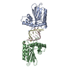
| Title | Structural Insights into HIV-1 Vif-Mediated Ubiquitination and Degradation of APOBEC3H | |||||||||
 Map data Map data | ||||||||||
 Sample Sample |
| |||||||||
 Keywords Keywords | APOBEC3H HIV-1 Vif / VIRAL PROTEIN / VIRAL PROTEIN-RNA complex | |||||||||
| Function / homology |  Function and homology information Function and homology informationRUNX3 regulates RUNX1-mediated transcription / RUNX1 regulates transcription of genes involved in BCR signaling / RUNX1 regulates transcription of genes involved in interleukin signaling / RUNX2 regulates bone development / core-binding factor complex / RUNX1 regulates expression of components of tight junctions / positive regulation of CD8-positive, alpha-beta T cell differentiation / RUNX2 regulates chondrocyte maturation / single-stranded DNA cytosine deaminase / negative regulation of CD4-positive, alpha-beta T cell differentiation ...RUNX3 regulates RUNX1-mediated transcription / RUNX1 regulates transcription of genes involved in BCR signaling / RUNX1 regulates transcription of genes involved in interleukin signaling / RUNX2 regulates bone development / core-binding factor complex / RUNX1 regulates expression of components of tight junctions / positive regulation of CD8-positive, alpha-beta T cell differentiation / RUNX2 regulates chondrocyte maturation / single-stranded DNA cytosine deaminase / negative regulation of CD4-positive, alpha-beta T cell differentiation / clearance of foreign intracellular DNA / cytidine deaminase activity / RUNX1 and FOXP3 control the development of regulatory T lymphocytes (Tregs) / lymphocyte differentiation / RUNX2 regulates genes involved in cell migration / Transcriptional regulation by RUNX2 / RUNX1 regulates transcription of genes involved in differentiation of keratinocytes / RUNX2 regulates genes involved in differentiation of myeloid cells / transposable element silencing / RUNX3 Regulates Immune Response and Cell Migration / myeloid cell differentiation / definitive hemopoiesis / target-directed miRNA degradation / elongin complex / RUNX1 regulates transcription of genes involved in differentiation of myeloid cells / Regulation of RUNX1 Expression and Activity / VCB complex / RUNX1 regulates transcription of genes involved in WNT signaling / RUNX1 regulates estrogen receptor mediated transcription / Cul5-RING ubiquitin ligase complex / negative regulation of viral genome replication / ubiquitin-dependent protein catabolic process via the C-end degron rule pathway / Cul2-RING ubiquitin ligase complex / RUNX2 regulates osteoblast differentiation / RUNX1 interacts with co-factors whose precise effect on RUNX1 targets is not known / Pausing and recovery of Tat-mediated HIV elongation / Tat-mediated HIV elongation arrest and recovery / HIV elongation arrest and recovery / Pausing and recovery of HIV elongation / RUNX3 regulates p14-ARF / Tat-mediated elongation of the HIV-1 transcript / Formation of HIV-1 elongation complex containing HIV-1 Tat / Formation of HIV elongation complex in the absence of HIV Tat / RNA Polymerase II Transcription Elongation / Formation of RNA Pol II elongation complex / viral life cycle / cell maturation / RNA Polymerase II Pre-transcription Events / transcription corepressor binding / TP53 Regulates Transcription of DNA Repair Genes / transcription initiation at RNA polymerase II promoter / transcription elongation by RNA polymerase II / RUNX1 regulates genes involved in megakaryocyte differentiation and platelet function / Regulation of RUNX3 expression and activity / Inactivation of CSF3 (G-CSF) signaling / Vif-mediated degradation of APOBEC3G / virion component / Evasion by RSV of host interferon responses / Oxygen-dependent proline hydroxylation of Hypoxia-inducible Factor Alpha / Transcriptional regulation of granulopoiesis / Regulation of expression of SLITs and ROBOs / protein polyubiquitination / osteoblast differentiation / Regulation of RUNX2 expression and activity / positive regulation of proteasomal ubiquitin-dependent protein catabolic process / Antigen processing: Ubiquitination & Proteasome degradation / RUNX1 regulates transcription of genes involved in differentiation of HSCs / Neddylation / protein-containing complex assembly / transcription by RNA polymerase II / Estrogen-dependent gene expression / sequence-specific DNA binding / defense response to virus / ubiquitin-dependent protein catabolic process / host cell cytoplasm / protein-macromolecule adaptor activity / transcription coactivator activity / protein ubiquitination / ubiquitin protein ligase binding / regulation of transcription by RNA polymerase II / host cell plasma membrane / negative regulation of transcription by RNA polymerase II / positive regulation of transcription by RNA polymerase II / RNA binding / zinc ion binding / nucleoplasm / membrane / cytoplasm / cytosol Similarity search - Function | |||||||||
| Biological species |   Homo sapiens (human) / Homo sapiens (human) /   Human immunodeficiency virus 1 / Human immunodeficiency virus 1 /  | |||||||||
| Method | single particle reconstruction / cryo EM / Resolution: 4.0 Å | |||||||||
 Authors Authors | Matsuo H / Skorupka KA | |||||||||
| Funding support |  United States, 1 items United States, 1 items
| |||||||||
 Citation Citation | Journal: Nat Commun / Year: 2025 Title: HIV-1 vif mediates ubiquitination of the proximal protomer in the APOBEC3H dimer to induce degradation. Authors: Katarzyna A Skorupka / Kazuhiro Matsuoka / Bakar Hassan / Rodolfo Ghirlando / Vanivilasini Balachandran / Ting-Hua Chen / Kylie J Walters / Celia A Schiffer / Matthias Wolf / Yasumasa ...Authors: Katarzyna A Skorupka / Kazuhiro Matsuoka / Bakar Hassan / Rodolfo Ghirlando / Vanivilasini Balachandran / Ting-Hua Chen / Kylie J Walters / Celia A Schiffer / Matthias Wolf / Yasumasa Iwatani / Hiroshi Matsuo /   Abstract: The APOBEC3 family of cytidine deaminases restricts retroviruses like HIV-1 by mutating viral DNA. HIV-1 evades this restriction by producing Vif, which recruits the Cullin-5 (CUL5) E3 ubiquitin ...The APOBEC3 family of cytidine deaminases restricts retroviruses like HIV-1 by mutating viral DNA. HIV-1 evades this restriction by producing Vif, which recruits the Cullin-5 (CUL5) E3 ubiquitin ligase complex to promote APOBEC3 degradation. Here we resolve key aspects of this counter-defense mechanism by determining a 3.6 Å cryo-EM structure of chimpanzee APOBEC3H (cpzA3H) in complex with HIV-1 Vif and three components of the CUL5 E3 ligase-CBFβ, EloB, and EloC (VCBC). The structure captures cpzA3H as an RNA-mediated dimer within the cpzA3H-VCBC complex, allowing us to examine the role of dimerization. We find that ubiquitination occurs specifically at two lysine residues on the Vif-proximal protomer, while the distal protomer remains unmodified. The structural model of the active cpzA3H-Vif-CUL5 E3 ligase holoenzyme reveals spatial preferences for ubiquitin transfer to the targeted lysine residues. These findings enhance our understanding of A3H degradation and suggest new antiviral strategies targeting this host-virus interface. | |||||||||
| History |
|
- Structure visualization
| Supplemental images |
|---|

- Downloads & links
-EMDB archive
| Map data | emd_47805.map.gz | 117.6 MB | EMDB map data format | |
|---|---|---|---|---|
| Header (meta data) | emd-47805-v30.xmlemd-47805.xml | 26.6 KB 26.6 KB | Display Display | EMDB header |
| FSC (resolution estimation) | emd_47805_fsc.xml | 13.2 KB | Display | FSC data file |
| Images |  emd_47805.png emd_47805.png | 80.5 KB | ||
| Filedesc metadata | emd-47805.cif.gz | 7.2 KB | ||
| Others | emd_47805_half_map_1.map.gzemd_47805_half_map_2.map.gz | 152 MB 152 MB | ||
| Archive directory |  http://ftp.pdbj.org/pub/emdb/structures/EMD-47805ftp://ftp.pdbj.org/pub/emdb/structures/EMD-47805 http://ftp.pdbj.org/pub/emdb/structures/EMD-47805ftp://ftp.pdbj.org/pub/emdb/structures/EMD-47805 | HTTPS FTP |
-Related structure data
| Related structure data |  9e9vMC  9e93C M: atomic model generated by this map C: citing same article ( |
|---|---|
| Similar structure data |

-Links
| EMDB pages | EMDB (EBI/PDBe) / EMDataResource |
|---|---|
| Related items in Molecule of the Month |


-Map
| File | Download / File: emd_47805.map.gz / Format: CCP4 / Size: 163.6 MB / Type: IMAGE STORED AS FLOATING POINT NUMBER (4 BYTES) | ||||||||||||||||||||||||||||||||||||
|---|---|---|---|---|---|---|---|---|---|---|---|---|---|---|---|---|---|---|---|---|---|---|---|---|---|---|---|---|---|---|---|---|---|---|---|---|---|
| Projections & slices | Image control
Images are generated by Spider. | ||||||||||||||||||||||||||||||||||||
| Voxel size | X=Y=Z: 0.81 Å | ||||||||||||||||||||||||||||||||||||
| Density |
| ||||||||||||||||||||||||||||||||||||
| Symmetry | Space group: 1 | ||||||||||||||||||||||||||||||||||||
| Details | EMDB XML:
|
 Z (Sec.)
 Y (Row.)
 X (Col.)


-Supplemental data
-Half map: #2
| File | emd_47805_half_map_1.map | ||||||||||||
|---|---|---|---|---|---|---|---|---|---|---|---|---|---|
| Projections & Slices |
| ||||||||||||
| Density Histograms |


-Half map: #1
| File | emd_47805_half_map_2.map | ||||||||||||
|---|---|---|---|---|---|---|---|---|---|---|---|---|---|
| Projections & Slices |
| ||||||||||||
| Density Histograms |


- Sample components
-Entire : Ternary complex of APOBEC3H and Vif of HIV-1
| Entire | Name: Ternary complex of APOBEC3H and Vif of HIV-1 |
|---|---|
| Components |
|
-Supramolecule #1: Ternary complex of APOBEC3H and Vif of HIV-1
| Supramolecule | Name: Ternary complex of APOBEC3H and Vif of HIV-1 / type: complex / ID: 1 / Parent: 0 / Macromolecule list: #2-#3 |
|---|---|
| Source (natural) | Organism: |
-Macromolecule #1: single-stranded DNA cytosine deaminase
| Macromolecule | Name: single-stranded DNA cytosine deaminase / type: protein_or_peptide / ID: 1 / Number of copies: 3 / Enantiomer: LEVO / EC number: single-stranded DNA cytosine deaminase |
|---|---|
| Source (natural) | Organism: |
| Molecular weight | Theoretical: 21.76818 KDa |
| Recombinant expression | Organism: |
| Sequence | String: MALLTAETFR LQFNNRRRLR RPYYPRKALL CYQLTPQNGS TPTRGYFENK KKCHAEICFI NEIKSMGLDE TQCYQVTCYL TWSPCSSCA WKLVDFIQAH DHLNLRIFAS RLYYHWCKPQ QEGLRLLCGS QVPVEVMGLP EFNDCWENFV DHEKPLSFDP C KMLEELDK NSRAIKRRLE RIKQS UniProtKB: single-stranded DNA cytosine deaminase |
-Macromolecule #4: Elongin-C
| Macromolecule | Name: Elongin-C / type: protein_or_peptide / ID: 4 / Number of copies: 1 / Enantiomer: LEVO |
|---|---|
| Source (natural) | Organism: Homo sapiens (human) |
| Molecular weight | Theoretical: 10.84342 KDa |
| Recombinant expression | Organism: |
| Sequence | String: MYVKLISSDG HEFIVKREHA LTSGTIKAML SGPGQFAENE TNEVNFREIP SHVLSKVCMY FTYKVRYTNS STEIPEFPIA PEIALELLM AANFLDC UniProtKB: Elongin-C |
-Macromolecule #5: Elongin-B
| Macromolecule | Name: Elongin-B / type: protein_or_peptide / ID: 5 / Number of copies: 1 / Enantiomer: LEVO |
|---|---|
| Source (natural) | Organism: Homo sapiens (human) |
| Molecular weight | Theoretical: 11.48803 KDa |
| Recombinant expression | Organism: |
| Sequence | String: MDVFLMIRRH KTTIFTDAKE SSTVFELKRI VEGILKRPPD EQRLYKDDQL LDDGKTLGEC GFTSQTARPQ APATVGLAFR ADDTFEALC IEPFSSPPEL PDV UniProtKB: Elongin-B |
-Macromolecule #6: Virion infectivity factor
| Macromolecule | Name: Virion infectivity factor / type: protein_or_peptide / ID: 6 / Number of copies: 2 / Enantiomer: LEVO |
|---|---|
| Source (natural) | Organism: Human immunodeficiency virus 1 |
| Molecular weight | Theoretical: 20.98224 KDa |
| Recombinant expression | Organism: |
| Sequence | String: MENRWQVMIV WQVDRMRINT WKRLVKHHMY ISRKAKDWFY RHHYESTNPK ISSEVHIPLG DAKLVITTYW GLHTGERDWH LGQGVSIEW RKKRYSTQVD PDLADQLIHL HYFDCFSESA IRNTILGRIV SPRCEYQAGH NKVGSLQYLA LAALIKPKQI K PPLPSVRK LTEDRWNK UniProtKB: Virion infectivity factor |
-Macromolecule #7: Core-binding factor subunit beta
| Macromolecule | Name: Core-binding factor subunit beta / type: protein_or_peptide / ID: 7 / Number of copies: 2 / Enantiomer: LEVO |
|---|---|
| Source (natural) | Organism: Homo sapiens (human) |
| Molecular weight | Theoretical: 20.272629 KDa |
| Recombinant expression | Organism: |
| Sequence | String: MPRVVPDQRS KFENEEFFRK LSRECEIKYT GFRDRPHEER QARFQNACRD GRSEIAFVAT GTNLSLQFFP ASWQGEQRQT PSREYVDLE REAGKVYLKA PMILNGVCVI WKGWIDLQRL DGMGCLEFDE ERAQQEDALA QQAFEEARRR TREFEDRDRS H REEMEVRV SQ UniProtKB: Core-binding factor subunit beta |
-Macromolecule #2: RNA (5'-R(P*CP*CP*CP*GP*GP*CP*A)-3')
| Macromolecule | Name: RNA (5'-R(P*CP*CP*CP*GP*GP*CP*A)-3') / type: rna / ID: 2 / Number of copies: 2 |
|---|---|
| Source (natural) | Organism: |
| Molecular weight | Theoretical: 3.159965 KDa |
| Sequence | String: AUACCCGGCA |
-Macromolecule #3: RNA (5'-R(P*GP*CP*CP*GP*GP*G)-3')
| Macromolecule | Name: RNA (5'-R(P*GP*CP*CP*GP*GP*G)-3') / type: rna / ID: 3 / Number of copies: 2 |
|---|---|
| Source (natural) | Organism: |
| Molecular weight | Theoretical: 2.887767 KDa |
| Sequence | String: UGCCGGGUA |
-Macromolecule #8: ZINC ION
| Macromolecule | Name: ZINC ION / type: ligand / ID: 8 / Number of copies: 4 / Formula: ZN |
|---|---|
| Molecular weight | Theoretical: 65.409 Da |
-Experimental details
-Structure determination
| Method | cryo EM |
|---|---|
 Processing Processing | single particle reconstruction |
| Aggregation state | particle |
-Sample preparation
| Concentration | 0.3 mg/mL | |||||||||||||||
|---|---|---|---|---|---|---|---|---|---|---|---|---|---|---|---|---|
| Buffer | pH: 7.4 Component:
| |||||||||||||||
| Vitrification | Cryogen name: ETHANE / Chamber humidity: 100 % / Chamber temperature: 281 K / Instrument: FEI VITROBOT MARK IV |
- Electron microscopy
| Microscope | FEI TECNAI ARCTICA |
|---|---|
| Image recording | Film or detector model: GATAN K3 (6k x 4k) / Number grids imaged: 3 / Number real images: 44142 / Average electron dose: 45.0 e/Å2 |
| Electron beam | Acceleration voltage: 200 kV / Electron source:  FIELD EMISSION GUN FIELD EMISSION GUN |
| Electron optics | C2 aperture diameter: 50.0 µm / Illumination mode: FLOOD BEAM / Imaging mode: BRIGHT FIELD / Cs: 2.7 mm / Nominal defocus max: 2.2 µm / Nominal defocus min: 0.8 µm |
| Sample stage | Cooling holder cryogen: NITROGEN |
| Experimental equipment |  Model: Talos Arctica / Image courtesy: FEI Company |
+Image processing

-Atomic model buiding 1
| Initial model |
| ||||||||||||||||||
|---|---|---|---|---|---|---|---|---|---|---|---|---|---|---|---|---|---|---|---|
| Refinement | Protocol: RIGID BODY FIT / Overall B value: 135 | ||||||||||||||||||
| Output model | PDB-9e9v: |

