[English] 日本語
Yorodumi
- EMDB-47493: Cryo-EM structure of the human P2X7 receptor in the UB-MBX-46-bou... -

+
Open data


ID or keywords:

Loading...

-
Basic information

Entry
Database: EMDB / ID: EMD-47493
TitleCryo-EM structure of the human P2X7 receptor in the UB-MBX-46-bound inhibited state
Map dataSharpened map for human P2X7 in the UB-MBX-46-bound inhibited state
Sample
  • Complex: Membrane protein
    • Protein or peptide: P2X purinoceptor 7
  • Ligand: (1s,3R,4R,6r,8S,9S)-N'-(2-chlorophenyl)-3,4,8,9-tetramethyltetracyclo[4.4.0.0~3,9~.0~4,8~]decane-1-carbohydrazide
  • Ligand: GUANOSINE-5'-DIPHOSPHATE
  • Ligand: ZINC ION
  • Ligand: 2-acetamido-2-deoxy-beta-D-glucopyranose
  • Ligand: CHOLESTEROL HEMISUCCINATE
  • Ligand: PALMITIC ACID
  • Ligand: SODIUM ION
  • Ligand: water
KeywordsMembrane Protein / Ion Channel / Ligand-Gated Ion Channel / P2X Receptor / P2XR / Allosteric Antagonist / Agonist
Function / homology
Function and homology information


positive regulation of bleb assembly / NAD transport / Platelet homeostasis / phagolysosome assembly / positive regulation of cytoskeleton organization / phospholipid transfer to membrane / positive regulation of monoatomic ion transmembrane transport / purinergic nucleotide receptor signaling pathway / gamma-aminobutyric acid secretion / extracellularly ATP-gated monoatomic cation channel activity ...positive regulation of bleb assembly / NAD transport / Platelet homeostasis / phagolysosome assembly / positive regulation of cytoskeleton organization / phospholipid transfer to membrane / positive regulation of monoatomic ion transmembrane transport / purinergic nucleotide receptor signaling pathway / gamma-aminobutyric acid secretion / extracellularly ATP-gated monoatomic cation channel activity / pore complex assembly / positive regulation of interleukin-1 alpha production / purinergic nucleotide receptor activity / positive regulation of gamma-aminobutyric acid secretion / bleb / Elevation of cytosolic Ca2+ levels / plasma membrane phospholipid scrambling / negative regulation of cell volume / collagen metabolic process / positive regulation of prostaglandin secretion / T cell apoptotic process / bleb assembly / response to fluid shear stress / mitochondrial depolarization / vesicle budding from membrane / ceramide biosynthetic process / positive regulation of T cell apoptotic process / prostaglandin secretion / cellular response to dsRNA / glutamate secretion / positive regulation of glutamate secretion / negative regulation of bone resorption / positive regulation of macrophage cytokine production / skeletal system morphogenesis / Mechanical load activates signaling by PIEZO1 and integrins in osteocytes / response to zinc ion / response to ATP / positive regulation of mitochondrial depolarization / positive regulation of NLRP3 inflammasome complex assembly / sodium channel activity / cellular response to ATP / protein homotrimerization / T cell homeostasis / monoatomic ion channel activity / positive regulation of calcium ion transport into cytosol / membrane protein ectodomain proteolysis / The NLRP3 inflammasome / protein secretion / synaptic vesicle exocytosis / response to electrical stimulus / membrane depolarization / potassium channel activity / positive regulation of bone mineralization / response to mechanical stimulus / T cell proliferation / Purinergic signaling in leishmaniasis infection / regulation of sodium ion transport / negative regulation of MAPK cascade / extrinsic apoptotic signaling pathway / release of sequestered calcium ion into cytosol / sensory perception of pain / homeostasis of number of cells within a tissue / reactive oxygen species metabolic process / positive regulation of glycolytic process / response to ischemia / positive regulation of interleukin-1 beta production / positive regulation of protein secretion / mitochondrion organization / neuromuscular junction / apoptotic signaling pathway / lipopolysaccharide binding / protein catabolic process / calcium-mediated signaling / T cell mediated cytotoxicity / response to calcium ion / calcium ion transmembrane transport / protein processing / positive regulation of interleukin-6 production / positive regulation of T cell mediated cytotoxicity / cell morphogenesis / cell-cell junction / presynapse / MAPK cascade / response to lipopolysaccharide / cell surface receptor signaling pathway / postsynapse / positive regulation of MAPK cascade / defense response to Gram-positive bacterium / response to xenobiotic stimulus / inflammatory response / signaling receptor binding / external side of plasma membrane / neuronal cell body / positive regulation of gene expression / mitochondrion / ATP binding / identical protein binding / membrane / plasma membrane / cytoplasm
Similarity search - Function
P2X purinoreceptor 7, intracellular domain / P2X purinoreceptor 7 intracellular domain / P2X7 purinoceptor / : / ATP P2X receptors signature. / ATP P2X receptor / P2X purinoreceptor / P2X purinoreceptor extracellular domain superfamily
Similarity search - Domain/homology
Biological speciesHomo sapiens (human)
Methodsingle particle reconstruction / cryo EM / Resolution: 2.53 Å
AuthorsOken AC / Turcu AL / Vazquez S / Mansoor SE
Funding support United States, 2 items
OrganizationGrant numberCountry
National Institutes of Health/National Heart, Lung, and Blood Institute (NIH/NHLBI)R00HL138129 United States
National Institutes of Health/National Institute of General Medical Sciences (NIH/NIGMS)DP2GM149551 United States
Citation
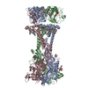
Journal: Nat Commun / Year: 2025
Title: A polycyclic scaffold identified by structure-based drug design effectively inhibits the human P2X7 receptor.
Authors: Adam C Oken / Andreea L Turcu / Eva Tzortzini / Kyriakos Georgiou / Jessica Nagel / Franka G Westermann / Marta Barniol-Xicota / Jonas Seidler / Ga-Ram Kim / So-Deok Lee / Annette Nicke / ...Authors: Adam C Oken / Andreea L Turcu / Eva Tzortzini / Kyriakos Georgiou / Jessica Nagel / Franka G Westermann / Marta Barniol-Xicota / Jonas Seidler / Ga-Ram Kim / So-Deok Lee / Annette Nicke / Yong-Chul Kim / Christa E Müller / Antonios Kolocouris / Santiago Vázquez / Steven E Mansoor /
Abstract: The P2X7 receptor is an ATP-gated ion channel that activates inflammatory pathways involved in diseases such as cancer, atherosclerosis, and neurodegeneration. However, despite the potential benefits ...The P2X7 receptor is an ATP-gated ion channel that activates inflammatory pathways involved in diseases such as cancer, atherosclerosis, and neurodegeneration. However, despite the potential benefits of blocking overactive signaling, no P2X7 receptor antagonists have been approved for clinical use. Understanding species-specific pharmacological effects of existing antagonists has been challenging, in part due to the dearth of molecular information on receptor orthologs. Here, to identify distinct molecular features in the human receptor, we determine high-resolution cryo-EM structures of the full-length wild-type human P2X7 receptor in apo closed and ATP-bound open state conformations and draw comparisons with structures of other orthologs. We also report a cryo-EM structure of the human receptor in complex with an adamantane-based inhibitor, which we leverage, in conjunction with functional data and molecular dynamics simulations, to design a potent and selective antagonist with a unique polycyclic scaffold. Functional and structural analysis reveal how this optimized ligand, termed UB-MBX-46, interacts with the classical allosteric pocket of the human P2X7 receptor with subnanomolar potency and high selectivity, revealing its significant therapeutic potential.
History
DepositionOct 23, 2024-
Header (metadata) releaseSep 24, 2025-
Map releaseSep 24, 2025-
UpdateSep 24, 2025-
Current statusSep 24, 2025Processing site: RCSB / Status: Released

-
Structure visualization

Supplemental images

Downloads & links

-
Map

FileDownload / File: emd_47493.map.gz / Format: CCP4 / Size: 824 MB / Type: IMAGE STORED AS FLOATING POINT NUMBER (4 BYTES)
AnnotationSharpened map for human P2X7 in the UB-MBX-46-bound inhibited state
Projections & slices

Image control

Size
Brightness
Contrast
Others
AxesZ (Sec.)Y (Row.)X (Col.)
0.65 Å/pix.
x 600 pix.
= 388.8 Å
0.65 Å/pix.
x 600 pix.
= 388.8 Å
0.65 Å/pix.
x 600 pix.
= 388.8 Å

Surface

Projections

Slices (1/3)

Slices (1/2)

Slices (2/3)

Images are generated by Spider.

Voxel sizeX=Y=Z: 0.648 Å
Density
Contour LevelBy AUTHOR: 0.15
Minimum - Maximum-0.88833606 - 1.2968866
Average (Standard dev.)0.00019092085 (±0.017386083)
SymmetrySpace group: 1
Details

EMDB XML:

Map geometry
Axis orderXYZ
Origin000
Dimensions600600600
Spacing600600600
CellA=B=C: 388.8 Å
α=β=γ: 90.0 °

-
Supplemental data

-
Mask #1

Fileemd_47493_msk_1.map
Projections & Slices
AxesZYX

Projections

Slices (1/2)
Density Histograms

-
Additional map: Unsharpened map for human P2X7 in the UB-MBX-46-bound inhibited state

Fileemd_47493_additional_1.map
AnnotationUnsharpened map for human P2X7 in the UB-MBX-46-bound inhibited state
Projections & Slices
AxesZYX

Projections

Slices (1/2)
Density Histograms

-
Half map: Half map A for human P2X7 in the UB-MBX-46-bound inhibited state

Fileemd_47493_half_map_1.map
AnnotationHalf map A for human P2X7 in the UB-MBX-46-bound inhibited state
Projections & Slices
AxesZYX

Projections

Slices (1/2)
Density Histograms

-
Half map: Half map B for human P2X7 in the UB-MBX-46-bound inhibited state

Fileemd_47493_half_map_2.map
AnnotationHalf map B for human P2X7 in the UB-MBX-46-bound inhibited state
Projections & Slices
AxesZYX

Projections

Slices (1/2)
Density Histograms

-
Sample components

+
Entire : Membrane protein

EntireName: Membrane protein
Components
  • Complex: Membrane protein
    • Protein or peptide: P2X purinoceptor 7
  • Ligand: (1s,3R,4R,6r,8S,9S)-N'-(2-chlorophenyl)-3,4,8,9-tetramethyltetracyclo[4.4.0.0~3,9~.0~4,8~]decane-1-carbohydrazide
  • Ligand: GUANOSINE-5'-DIPHOSPHATE
  • Ligand: ZINC ION
  • Ligand: 2-acetamido-2-deoxy-beta-D-glucopyranose
  • Ligand: CHOLESTEROL HEMISUCCINATE
  • Ligand: PALMITIC ACID
  • Ligand: SODIUM ION
  • Ligand: water

+
Supramolecule #1: Membrane protein

SupramoleculeName: Membrane protein / type: complex / ID: 1 / Parent: 0 / Macromolecule list: #1
Source (natural)Organism: Homo sapiens (human)

+
Macromolecule #1: P2X purinoceptor 7

MacromoleculeName: P2X purinoceptor 7 / type: protein_or_peptide / ID: 1 / Number of copies: 3 / Enantiomer: LEVO
Source (natural)Organism: Homo sapiens (human)
Molecular weightTheoretical: 68.67182 KDa
Recombinant expressionOrganism: Homo sapiens (human)
SequenceString: MPACCSCSDV FQYETNKVTR IQSMNYGTIK WFFHVIIFSY VCFALVSDKL YQRKEPVISS VHTKVKGIAE VKEEIVENGV KKLVHSVFD TADYTFPLQG NSFFVMTNFL KTEGQEQRLC PEYPTRRTLC SSDRGCKKGW MDPQSKGIQT GRCVVYEGNQ K TCEVSAWC ...String:
MPACCSCSDV FQYETNKVTR IQSMNYGTIK WFFHVIIFSY VCFALVSDKL YQRKEPVISS VHTKVKGIAE VKEEIVENGV KKLVHSVFD TADYTFPLQG NSFFVMTNFL KTEGQEQRLC PEYPTRRTLC SSDRGCKKGW MDPQSKGIQT GRCVVYEGNQ K TCEVSAWC PIEAVEEAPR PALLNSAENF TVLIKNNIDF PGHNYTTRNI LPGLNITCTF HKTQNPQCPI FRLGDIFRET GD NFSDVAI QGGIMGIEIY WDCNLDRWFH HCRPKYSFRR LDDKTTNVSL YPGYNFRYAK YYKENNVEKR TLIKVFGIRF DIL VFGTGG KFDIIQLVVY IGSTLSYFGL AAVFIDFLID TYSSNCCRSH IYPWCKCCQP CVVNEYYYRK KCESIVEPKP TLKY VSFVD ESHIRMVNQQ LLGRSLQDVK GQEVPRPAMD FTDLSRLPLA LHDTPPIPGQ PEEIQLLRKE ATPRSRDSPV WCQCG SCLP SQLPESHRCL EELCCRKKPG ACITTSELFR KLVLSRHVLQ FLLLYQEPLL ALDVDSTNSR LRHCAYRCYA TWRFGS QDM ADFAILPSCC RWRIRKEFPK SEGQYSGFKS PY

UniProtKB: P2X purinoceptor 7

+
Macromolecule #3: (1s,3R,4R,6r,8S,9S)-N'-(2-chlorophenyl)-3,4,8,9-tetramethyltetrac...

MacromoleculeName: (1s,3R,4R,6r,8S,9S)-N'-(2-chlorophenyl)-3,4,8,9-tetramethyltetracyclo[4.4.0.0~3,9~.0~4,8~]decane-1-carbohydrazide
type: ligand / ID: 3 / Number of copies: 3 / Formula: A1BD9
Molecular weightTheoretical: 358.905 Da

+
Macromolecule #4: GUANOSINE-5'-DIPHOSPHATE

MacromoleculeName: GUANOSINE-5'-DIPHOSPHATE / type: ligand / ID: 4 / Number of copies: 3 / Formula: GDP
Molecular weightTheoretical: 443.201 Da
Chemical component information

ChemComp-GDP:
GUANOSINE-5'-DIPHOSPHATE / GDP, energy-carrying molecule*YM

+
Macromolecule #5: ZINC ION

MacromoleculeName: ZINC ION / type: ligand / ID: 5 / Number of copies: 6 / Formula: ZN
Molecular weightTheoretical: 65.409 Da

+
Macromolecule #6: 2-acetamido-2-deoxy-beta-D-glucopyranose

MacromoleculeName: 2-acetamido-2-deoxy-beta-D-glucopyranose / type: ligand / ID: 6 / Number of copies: 6 / Formula: NAG
Molecular weightTheoretical: 221.208 Da
Chemical component information

ChemComp-NAG:
2-acetamido-2-deoxy-beta-D-glucopyranose

+
Macromolecule #7: CHOLESTEROL HEMISUCCINATE

MacromoleculeName: CHOLESTEROL HEMISUCCINATE / type: ligand / ID: 7 / Number of copies: 6 / Formula: Y01
Molecular weightTheoretical: 486.726 Da
Chemical component information

ChemComp-Y01:
CHOLESTEROL HEMISUCCINATE

+
Macromolecule #8: PALMITIC ACID

MacromoleculeName: PALMITIC ACID / type: ligand / ID: 8 / Number of copies: 21 / Formula: PLM
Source (natural)Organism: Homo sapiens (human)
Molecular weightTheoretical: 256.424 Da
Chemical component information

ChemComp-PLM:
PALMITIC ACID

+
Macromolecule #9: SODIUM ION

MacromoleculeName: SODIUM ION / type: ligand / ID: 9 / Number of copies: 1
Molecular weightTheoretical: 22.99 Da

+
Macromolecule #10: water

MacromoleculeName: water / type: ligand / ID: 10 / Number of copies: 273 / Formula: HOH
Molecular weightTheoretical: 18.015 Da
Chemical component information

ChemComp-HOH:
WATER

-
Experimental details

-
Structure determination

Methodcryo EM
Processingsingle particle reconstruction
Aggregation stateparticle

-
Sample preparation

BufferpH: 7
VitrificationCryogen name: ETHANE / Instrument: FEI VITROBOT MARK IV

-
Electron microscopy

MicroscopeFEI TITAN KRIOS
Specialist opticsEnergy filter - Slit width: 20 eV
Image recordingFilm or detector model: GATAN K3 BIOQUANTUM (6k x 4k) / Number real images: 24191 / Average electron dose: 42.0 e/Å2
Electron beamAcceleration voltage: 300 kV / Electron source: FIELD EMISSION GUN
Electron opticsC2 aperture diameter: 50.0 µm / Illumination mode: FLOOD BEAM / Imaging mode: BRIGHT FIELD / Cs: 2.7 mm / Nominal defocus max: 1.4000000000000001 µm / Nominal defocus min: 0.9 µm / Nominal magnification: 130000
Sample stageSpecimen holder model: FEI TITAN KRIOS AUTOGRID HOLDER
Experimental equipment
Model: Titan Krios / Image courtesy: FEI Company

+
Image processing

CTF correctionType: PHASE FLIPPING AND AMPLITUDE CORRECTION
Startup modelType of model: PDB ENTRY
PDB model - PDB ID:
Final reconstructionApplied symmetry - Point group: C3 (3 fold cyclic) / Resolution.type: BY AUTHOR / Resolution: 2.53 Å / Resolution method: FSC 0.143 CUT-OFF / Number images used: 190425
Initial angle assignmentType: ANGULAR RECONSTITUTION
Final angle assignmentType: ANGULAR RECONSTITUTION
FSC plot (resolution estimation)

+
About Yorodumi

-
News

-
Feb 9, 2022. New format data for meta-information of EMDB entries

New format data for meta-information of EMDB entries

  • Version 3 of the EMDB header file is now the official format.
  • The previous official version 1.9 will be removed from the archive.

Related info.:EMDB header

External links:wwPDB to switch to version 3 of the EMDB data model

-
Aug 12, 2020. Covid-19 info

Covid-19 info

URL: https://pdbj.org/emnavi/covid19.php

New page: Covid-19 featured information page in EM Navigator.

Related info.:Covid-19 info / Mar 5, 2020. Novel coronavirus structure data

+
Mar 5, 2020. Novel coronavirus structure data

Novel coronavirus structure data

Related info.:Yorodumi Speices / Aug 12, 2020. Covid-19 info

External links:COVID-19 featured content - PDBj / Molecule of the Month (242):Coronavirus Proteases

+
Jan 31, 2019. EMDB accession codes are about to change! (news from PDBe EMDB page)

EMDB accession codes are about to change! (news from PDBe EMDB page)

  • The allocation of 4 digits for EMDB accession codes will soon come to an end. Whilst these codes will remain in use, new EMDB accession codes will include an additional digit and will expand incrementally as the available range of codes is exhausted. The current 4-digit format prefixed with “EMD-” (i.e. EMD-XXXX) will advance to a 5-digit format (i.e. EMD-XXXXX), and so on. It is currently estimated that the 4-digit codes will be depleted around Spring 2019, at which point the 5-digit format will come into force.
  • The EM Navigator/Yorodumi systems omit the EMD- prefix.

Related info.:Q: What is EMD? / ID/Accession-code notation in Yorodumi/EM Navigator

External links:EMDB Accession Codes are Changing Soon! / Contact to PDBj

+
Jul 12, 2017. Major update of PDB

Major update of PDB

  • wwPDB released updated PDB data conforming to the new PDBx/mmCIF dictionary.
  • This is a major update changing the version number from 4 to 5, and with Remediation, in which all the entries are updated.
  • In this update, many items about electron microscopy experimental information are reorganized (e.g. em_software).
  • Now, EM Navigator and Yorodumi are based on the updated data.

External links:wwPDB Remediation / Enriched Model Files Conforming to OneDep Data Standards Now Available in the PDB FTP Archive

-
Yorodumi

Thousand views of thousand structures

  • Yorodumi is a browser for structure data from EMDB, PDB, SASBDB, etc.
  • This page is also the successor to EM Navigator detail page, and also detail information page/front-end page for Omokage search.
  • The word "yorodu" (or yorozu) is an old Japanese word meaning "ten thousand". "mi" (miru) is to see.

Related info.:EMDB / PDB / SASBDB / Comparison of 3 databanks / Yorodumi Search / Aug 31, 2016. New EM Navigator & Yorodumi / Yorodumi Papers / Jmol/JSmol / Function and homology information / Changes in new EM Navigator and Yorodumi

Read more