 Movie
Movie Controller

[English] 日本語
 Yorodumi
- EMDB-47476: Cryo-EM structure of PRMT5/WDR77 in complex with 6S complex (pICl... -
+ Open data

- Basic information
| Entry | 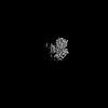 | |||||||||
|---|---|---|---|---|---|---|---|---|---|---|
| Title | Cryo-EM structure of PRMT5/WDR77 in complex with 6S complex (pICln PBM local refinement) | |||||||||
 Map data Map data | Map of locally refined PRMT5/WDR77/pICln PBM peptide | |||||||||
 Sample Sample |
| |||||||||
 Keywords Keywords | PRMT5 / Methyl transferase / WDR77 / arginine / TRANSFERASE | |||||||||
| Function / homology |  Function and homology information Function and homology informationpositive regulation of adenylate cyclase-inhibiting dopamine receptor signaling pathway / peptidyl-arginine N-methylation / oocyte axis specification / type II protein arginine methyltransferase / protein-arginine omega-N symmetric methyltransferase activity / peptidyl-arginine methylation / Golgi ribbon formation / negative regulation of epithelial cell proliferation involved in prostate gland development / secretory columnal luminar epithelial cell differentiation involved in prostate glandular acinus development / histone H4R3 methyltransferase activity ...positive regulation of adenylate cyclase-inhibiting dopamine receptor signaling pathway / peptidyl-arginine N-methylation / oocyte axis specification / type II protein arginine methyltransferase / protein-arginine omega-N symmetric methyltransferase activity / peptidyl-arginine methylation / Golgi ribbon formation / negative regulation of epithelial cell proliferation involved in prostate gland development / secretory columnal luminar epithelial cell differentiation involved in prostate glandular acinus development / histone H4R3 methyltransferase activity / : / epithelial cell proliferation involved in prostate gland development / protein-arginine N-methyltransferase activity / methylosome / pICln-Sm protein complex / positive regulation of mRNA splicing, via spliceosome / methyl-CpG binding / mRNA cis splicing, via spliceosome / cell volume homeostasis / chloride transport / endothelial cell activation / histone H3 methyltransferase activity / histone methyltransferase activity / regulation of mitotic nuclear division / negative regulation of gene expression via chromosomal CpG island methylation / Cul4B-RING E3 ubiquitin ligase complex / E-box binding / histone methyltransferase complex / positive regulation of oligodendrocyte differentiation / negative regulation of cell differentiation / spliceosomal snRNP assembly / ribonucleoprotein complex binding / regulation of ERK1 and ERK2 cascade / ubiquitin-like ligase-substrate adaptor activity / liver regeneration / regulation of signal transduction by p53 class mediator / methyltransferase activity / spliceosomal complex / circadian regulation of gene expression / DNA-templated transcription termination / Regulation of TP53 Activity through Methylation / RMTs methylate histone arginines / protein polyubiquitination / p53 binding / transcription corepressor activity / snRNP Assembly / ubiquitin-dependent protein catabolic process / transcription coactivator activity / cytoskeleton / cilium / chromatin remodeling / protein heterodimerization activity / intracellular membrane-bounded organelle / positive regulation of cell population proliferation / regulation of DNA-templated transcription / regulation of transcription by RNA polymerase II / chromatin / Golgi apparatus / RNA binding / nucleoplasm / identical protein binding / nucleus / plasma membrane / cytosol / cytoplasm Similarity search - Function | |||||||||
| Biological species |  Homo sapiens (human) Homo sapiens (human) | |||||||||
| Method | single particle reconstruction / cryo EM / Resolution: 3.36 Å | |||||||||
 Authors Authors | Jin CY / Hunkeler M / Fischer ES | |||||||||
| Funding support |  United States, 1 items United States, 1 items
| |||||||||
 Citation Citation | Journal: J Biol Chem / Year: 2025 Title: Substrate adaptors are flexible tethering modules that enhance substrate methylation by the arginine methyltransferase PRMT5. Authors: Cyrus Y Jin / Moritz Hunkeler / Kathleen M Mulvaney / William R Sellers / Eric S Fischer / Abstract: Protein arginine methyltransferase (PRMT) 5 is an essential arginine methyltransferase responsible for the majority of cellular symmetric dimethyl-arginine marks. PRMT5 uses substrate adaptors such ...Protein arginine methyltransferase (PRMT) 5 is an essential arginine methyltransferase responsible for the majority of cellular symmetric dimethyl-arginine marks. PRMT5 uses substrate adaptors such as pICln, RIOK1, and COPR5 to recruit and methylate a wide range of substrates. Although the substrate adaptors play important roles in substrate recognition, how they direct PRMT5 activity towards specific substrates remains incompletely understood. Using biochemistry and cryogenic electron microscopy, we show that these adaptors compete for the same binding site on PRMT5. We find that substrate adaptor and substrate complexes are bound to PRMT5 through two peptide motifs, enabling these adaptors to act as flexible tethering modules to enhance substrate methylation. Taken together, our results shed structural and mechanistic light on the PRMT5 substrate adaptor function and the biochemical nature of PRMT5 interactors. | |||||||||
| History |
|
- Structure visualization
| Supplemental images |
|---|

- Downloads & links
-EMDB archive
| Map data | emd_47476.map.gz | 52.1 MB | EMDB map data format | |
|---|---|---|---|---|
| Header (meta data) | emd-47476-v30.xmlemd-47476.xml | 23.3 KB 23.3 KB | Display Display | EMDB header |
| FSC (resolution estimation) | emd_47476_fsc.xml | 11.2 KB | Display | FSC data file |
| Images |  emd_47476.png emd_47476.png | 52.1 KB | ||
| Masks | emd_47476_msk_1.map | 103 MB | Mask map | |
| Filedesc metadata | emd-47476.cif.gz | 7.2 KB | ||
| Others | emd_47476_half_map_1.map.gzemd_47476_half_map_2.map.gz | 95.5 MB 95.5 MB | ||
| Archive directory |  http://ftp.pdbj.org/pub/emdb/structures/EMD-47476ftp://ftp.pdbj.org/pub/emdb/structures/EMD-47476 http://ftp.pdbj.org/pub/emdb/structures/EMD-47476ftp://ftp.pdbj.org/pub/emdb/structures/EMD-47476 | HTTPS FTP |
-Validation report
| Summary document | emd_47476_validation.pdf.gz | 825.4 KB | Display | EMDB validaton report |
|---|---|---|---|---|
| Full document | emd_47476_full_validation.pdf.gz | 824.9 KB | Display | |
| Data in XML | emd_47476_validation.xml.gz | 18.2 KB | Display | |
| Data in CIF | emd_47476_validation.cif.gz | 23.6 KB | Display | |
| Arichive directory | https://ftp.pdbj.org/pub/emdb/validation_reports/EMD-47476ftp://ftp.pdbj.org/pub/emdb/validation_reports/EMD-47476 | HTTPS FTP |
-Related structure data
| Related structure data |  9e3aMC  9e3bC  9e3cC M: atomic model generated by this map C: citing same article ( |
|---|---|
| Similar structure data |

-Links
| EMDB pages | EMDB (EBI/PDBe) / EMDataResource |
|---|---|
| Related items in Molecule of the Month |

-Map
| File | Download / File: emd_47476.map.gz / Format: CCP4 / Size: 103 MB / Type: IMAGE STORED AS FLOATING POINT NUMBER (4 BYTES) | ||||||||||||||||||||||||||||||||||||
|---|---|---|---|---|---|---|---|---|---|---|---|---|---|---|---|---|---|---|---|---|---|---|---|---|---|---|---|---|---|---|---|---|---|---|---|---|---|
| Annotation | Map of locally refined PRMT5/WDR77/pICln PBM peptide | ||||||||||||||||||||||||||||||||||||
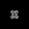
| Projections & slices | Image control
Images are generated by Spider. | ||||||||||||||||||||||||||||||||||||
| Voxel size | X=Y=Z: 1.408 Å | ||||||||||||||||||||||||||||||||||||
| Density |
| ||||||||||||||||||||||||||||||||||||
| Symmetry | Space group: 1 | ||||||||||||||||||||||||||||||||||||
| Details | EMDB XML:
|
 Z (Sec.)
 Y (Row.)
 X (Col.)


-Supplemental data
-Mask #1
| File | emd_47476_msk_1.map | ||||||||||||
|---|---|---|---|---|---|---|---|---|---|---|---|---|---|
| Projections & Slices |
| ||||||||||||
| Density Histograms |


-Half map: Half map of local refinement of PRMT5/WDR77/pICln PBM peptide
| File | emd_47476_half_map_1.map | ||||||||||||
|---|---|---|---|---|---|---|---|---|---|---|---|---|---|
| Annotation | Half map of local refinement of PRMT5/WDR77/pICln PBM peptide | ||||||||||||
| Projections & Slices |
| ||||||||||||
| Density Histograms |


-Half map: Half map of local refinement of PRMT5/WDR77/pICln PBM peptide
| File | emd_47476_half_map_2.map | ||||||||||||
|---|---|---|---|---|---|---|---|---|---|---|---|---|---|
| Annotation | Half map of local refinement of PRMT5/WDR77/pICln PBM peptide | ||||||||||||
| Projections & Slices |
| ||||||||||||
| Density Histograms |


- Sample components
-Entire : PRMT5/WDR77 in complex with 6S (PBM local refinement)
| Entire | Name: PRMT5/WDR77 in complex with 6S (PBM local refinement) |
|---|---|
| Components |
|
-Supramolecule #1: PRMT5/WDR77 in complex with 6S (PBM local refinement)
| Supramolecule | Name: PRMT5/WDR77 in complex with 6S (PBM local refinement) / type: complex / ID: 1 / Parent: 0 / Macromolecule list: all |
|---|---|
| Molecular weight | Theoretical: 770 KDa |
-Supramolecule #2: PRMT5/WDR77
| Supramolecule | Name: PRMT5/WDR77 / type: complex / ID: 2 / Parent: 1 / Macromolecule list: #1-#2 |
|---|---|
| Source (natural) | Organism: Homo sapiens (human) |
-Supramolecule #3: 6S complex
| Supramolecule | Name: 6S complex / type: complex / ID: 3 / Parent: 1 / Macromolecule list: #3 |
|---|---|
| Source (natural) | Organism: Homo sapiens (human) |
-Supramolecule #4: Methylosome subunit pICln
| Supramolecule | Name: Methylosome subunit pICln / type: complex / ID: 4 / Parent: 3 / Macromolecule list: #3 |
|---|
-Supramolecule #5: small nuclear ribonucleoproteins Sm D1, E, F, G
| Supramolecule | Name: small nuclear ribonucleoproteins Sm D1, E, F, G / type: complex / ID: 5 / Parent: 3 |
|---|
-Macromolecule #1: Protein arginine N-methyltransferase 5
| Macromolecule | Name: Protein arginine N-methyltransferase 5 / type: protein_or_peptide / ID: 1 / Number of copies: 1 / Enantiomer: LEVO / EC number: type II protein arginine methyltransferase |
|---|---|
| Source (natural) | Organism: Homo sapiens (human) |
| Molecular weight | Theoretical: 72.766664 KDa |
| Recombinant expression | Organism:  Trichoplusia ni (cabbage looper) Trichoplusia ni (cabbage looper) |
| Sequence | String: MAAMAVGGAG GSRVSSGRDL NCVPEIADTL GAVAKQGFDF LCMPVFHPRF KREFIQEPAK NRPGPQTRSD LLLSGRDWNT LIVGKLSPW IRPDSKVEKI RRNSEAAMLQ ELNFGAYLGL PAFLLPLNQE DNTNLARVLT NHIHTGHHSS MFWMRVPLVA P EDLRDDII ...String: MAAMAVGGAG GSRVSSGRDL NCVPEIADTL GAVAKQGFDF LCMPVFHPRF KREFIQEPAK NRPGPQTRSD LLLSGRDWNT LIVGKLSPW IRPDSKVEKI RRNSEAAMLQ ELNFGAYLGL PAFLLPLNQE DNTNLARVLT NHIHTGHHSS MFWMRVPLVA P EDLRDDII ENAPTTHTEE YSGEEKTWMW WHNFRTLCDY SKRIAVALEI GADLPSNHVI DRWLGEPIKA AILPTSIFLT NK KGFPVLS KMHQRLIFRL LKLEVQFIIT GTNHHSEKEF CSYLQYLEYL SQNRPPPNAY ELFAKGYEDY LQSPLQPLMD NLE SQTYEV FEKDPIKYSQ YQQAIYKCLL DRVPEEEKDT NVQVLMVLGA GRGPLVNASL RAAKQADRRI KLYAVEKNPN AVVT LENWQ FEEWGSQVTV VSSDMREWVA PEKADIIVSE LLGSFADNEL SPECLDGAQH FLKDDGVSIP GEYTSFLAPI SSSKL YNEV RACREKDRDP EAQFEMPYVV RLHNFHQLSA PQPCFTFSHP NRDPMIDNNR YCTLEFPVEV NTVLHGFAGY FETVLY QDI TLSIRPETHS PGMFSWFPIL FPIKQPITVR EGQTICVRFW RCSNSKKVWY EWAVTAPVCS AIHNPTGRSY TIGL UniProtKB: Protein arginine N-methyltransferase 5 |
-Macromolecule #2: Methylosome protein WDR77
| Macromolecule | Name: Methylosome protein WDR77 / type: protein_or_peptide / ID: 2 / Number of copies: 1 / Enantiomer: LEVO |
|---|---|
| Source (natural) | Organism: Homo sapiens (human) |
| Molecular weight | Theoretical: 37.186695 KDa |
| Recombinant expression | Organism: Trichoplusia ni (cabbage looper) |
| Sequence | String: GGGRTMRKET PPPLVPPAAR EWNLPPNAPA CMERQLEAAR YRSDGALLLG ASSLSGRCWA GSLWLFKDPC AAPNEGFCSA GVQTEAGVA DLTWVGERGI LVASDSGAVE LWELDENETL IVSKFCKYEH DDIVSTVSVL SSGTQAVSGS KDICIKVWDL A QQVVLSSY ...String: GGGRTMRKET PPPLVPPAAR EWNLPPNAPA CMERQLEAAR YRSDGALLLG ASSLSGRCWA GSLWLFKDPC AAPNEGFCSA GVQTEAGVA DLTWVGERGI LVASDSGAVE LWELDENETL IVSKFCKYEH DDIVSTVSVL SSGTQAVSGS KDICIKVWDL A QQVVLSSY RAHAAQVTCV AASPHKDSVF LSCSEDNRIL LWDTRCPKPA SQIGCSAPGY LPTSLAWHPQ QSEVFVFGDE NG TVSLVDT KSTSCVLSSA VHSQCVTGLV FSPHSVPFLA SLSEDCSLAV LDSSLSELFR SQAHRDFVRD ATWSPLNHSL LTT VGWDHQ VVHHVVPTEP LPAPGPASVT E UniProtKB: Methylosome protein WDR77 |
-Macromolecule #3: Methylosome subunit pICln
| Macromolecule | Name: Methylosome subunit pICln / type: protein_or_peptide / ID: 3 / Number of copies: 1 / Enantiomer: LEVO |
|---|---|
| Source (natural) | Organism: Homo sapiens (human) |
| Molecular weight | Theoretical: 26.552504 KDa |
| Recombinant expression | Organism:  |
| Sequence | String: SSGSMSFLKS FPPPGPAEGL LRQQPDTEAV LNGKGLGTGT LYIAESRLSW LDGSGLGFSL EYPTISLHAL SRDRSDCLGE HLYVMVNAK FEEESKEPVA DEEEEDSDDD VEPITEFRFV PSDKSALEAM FTAMCECQAL HPDPEDEDSD DYDGEEYDVE A HEQGQGDI ...String: SSGSMSFLKS FPPPGPAEGL LRQQPDTEAV LNGKGLGTGT LYIAESRLSW LDGSGLGFSL EYPTISLHAL SRDRSDCLGE HLYVMVNAK FEEESKEPVA DEEEEDSDDD VEPITEFRFV PSDKSALEAM FTAMCECQAL HPDPEDEDSD DYDGEEYDVE A HEQGQGDI PTFYTYEEGL SHLTAEGQAT LERLEGMLSQ SVSSQYNMAG VRTEDSIRDY EDGMEVDTTP TVAGQFEDAD VD H UniProtKB: Methylosome subunit pICln |
-Experimental details
-Structure determination
| Method | cryo EM |
|---|---|
 Processing Processing | single particle reconstruction |
| Aggregation state | particle |
-Sample preparation
| Concentration | 1 mg/mL | |||||||||||||||
|---|---|---|---|---|---|---|---|---|---|---|---|---|---|---|---|---|
| Buffer | pH: 7.4 Component:
Details: 30 mM HEPES pH 7.4, 150 mM NaCl, 3 mM TCEP, 3.33 mM sinefungin | |||||||||||||||
| Grid | Model: Quantifoil R1.2/1.3 / Material: COPPER / Mesh: 300 / Support film - Material: CARBON / Support film - topology: HOLEY ARRAY | |||||||||||||||
| Vitrification | Cryogen name: ETHANE / Chamber humidity: 90 % / Chamber temperature: 283 K / Instrument: LEICA EM GP |
- Electron microscopy
| Microscope | TFS TALOS |
|---|---|
| Image recording | Film or detector model: GATAN K3 (6k x 4k) / Number grids imaged: 1 / Number real images: 3615 / Average exposure time: 5.0 sec. / Average electron dose: 53.112 e/Å2 |
| Electron beam | Acceleration voltage: 200 kV / Electron source:  FIELD EMISSION GUN FIELD EMISSION GUN |
| Electron optics | C2 aperture diameter: 50.0 µm / Illumination mode: FLOOD BEAM / Imaging mode: BRIGHT FIELD / Cs: 2.7 mm / Nominal defocus max: 2.2 µm / Nominal defocus min: 0.8 µm / Nominal magnification: 36000 |
| Sample stage | Specimen holder model: FEI TITAN KRIOS AUTOGRID HOLDER / Cooling holder cryogen: NITROGEN |
+Image processing

-Atomic model buiding 1
| Initial model |
| ||||||||
|---|---|---|---|---|---|---|---|---|---|
| Refinement | Space: REAL / Protocol: OTHER / Overall B value: 98.6 / Target criteria: real space correlation | ||||||||
| Output model | PDB-9e3a: |

