 Movie
Movie Controller

[English] 日本語
 Yorodumi
- EMDB-47240: Gag CA-SP1 immature lattice bound with Lenacapavir from enveloped... -
+ Open data

- Basic information
| Entry | 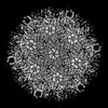 | |||||||||
|---|---|---|---|---|---|---|---|---|---|---|
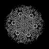
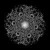
| Title | Gag CA-SP1 immature lattice bound with Lenacapavir from enveloped virus like particles (T8I) | |||||||||
 Map data Map data | anisotropic sharpened map | |||||||||
 Sample Sample |
| |||||||||
 Keywords Keywords | HIV-1 / Gag / CA-SP1 / Inhibitor / virion assembly / VIRAL PROTEIN | |||||||||
| Function / homology |  Function and homology information Function and homology informationSynthesis And Processing Of GAG, GAGPOL Polyproteins / host cellular component / host cell nuclear membrane / Integration of viral DNA into host genomic DNA / Autointegration results in viral DNA circles / Minus-strand DNA synthesis / Plus-strand DNA synthesis / Uncoating of the HIV Virion / 2-LTR circle formation / Vpr-mediated nuclear import of PICs ...Synthesis And Processing Of GAG, GAGPOL Polyproteins / host cellular component / host cell nuclear membrane / Integration of viral DNA into host genomic DNA / Autointegration results in viral DNA circles / Minus-strand DNA synthesis / Plus-strand DNA synthesis / Uncoating of the HIV Virion / 2-LTR circle formation / Vpr-mediated nuclear import of PICs / Early Phase of HIV Life Cycle / Integration of provirus / viral budding via host ESCRT complex / APOBEC3G mediated resistance to HIV-1 infection / Binding and entry of HIV virion / Membrane binding and targetting of GAG proteins / Assembly Of The HIV Virion / Budding and maturation of HIV virion / host multivesicular body / viral nucleocapsid / viral translational frameshifting / host cell plasma membrane / virion membrane / structural molecule activity / RNA binding / zinc ion binding Similarity search - Function | |||||||||
| Biological species |  Human immunodeficiency virus type 1 (NEW YORK-5 ISOLATE) Human immunodeficiency virus type 1 (NEW YORK-5 ISOLATE) | |||||||||
| Method | single particle reconstruction / cryo EM / Resolution: 3.13 Å | |||||||||
 Authors Authors | Wu C / Meuser ME / Xiong Y | |||||||||
| Funding support |  United States, 2 items United States, 2 items
| |||||||||
 Citation Citation | Journal: bioRxiv / Year: 2024 Title: Structural insights into inhibitor mechanisms on immature HIV-1 Gag lattice revealed by high-resolution single-particle cryo-EM. Authors: Chunxiang Wu / Megan E Meuser / Juan S Rey / Hamed Meshkin / Rachel Yang / Swapnil Chandrakant Devarkar / Christian Freniere / Jiong Shi / Christopher Aiken / Juan R Perilla / Yong Xiong / Abstract: HIV-1 inhibitors, such as Bevirimat (BVM) and Lenacapavir (LEN), block the production and maturation of infectious virions. However, their mechanisms remain unclear due to the absence of high- ...HIV-1 inhibitors, such as Bevirimat (BVM) and Lenacapavir (LEN), block the production and maturation of infectious virions. However, their mechanisms remain unclear due to the absence of high-resolution structures for BVM complexes and LEN's structural data being limited to the mature capsid. Utilizing perforated virus-like particles (VLPs) produced from mammalian cells, we developed an approach to determine cryo-electron microscopy (cryo-EM) structures of HIV-1 with inhibitors. This allowed for the first structural determination of the native immature HIV-1 particle with BVM and LEN bound inside the VLPs at high resolutions. Our findings offer a more accurate model of BVM engaging the Gag lattice and, importantly, demonstrate that LEN not only binds the mature capsid but also targets the immature lattice in a distinct manner. The binding of LEN induces a conformational change in the capsid protein (CA) region and alters the architecture of the Gag lattice, which may affect the maturation process. These insights expand our understanding of the inhibitory mechanisms of BVM and LEN on HIV-1 and provide valuable clues for the design of future inhibitors. | |||||||||
| History |
|
- Structure visualization
| Supplemental images |
|---|

- Downloads & links
-EMDB archive
| Map data | emd_47240.map.gz | 147.4 MB | EMDB map data format | |
|---|---|---|---|---|
| Header (meta data) | emd-47240-v30.xmlemd-47240.xml | 25.1 KB 25.1 KB | Display Display | EMDB header |
| FSC (resolution estimation) | emd_47240_fsc.xml | 11.5 KB | Display | FSC data file |
| Images |  emd_47240.png emd_47240.png | 165.9 KB | ||
| Masks | emd_47240_msk_1.map | 158 MB | Mask map | |
| Filedesc metadata | emd-47240.cif.gz | 7.4 KB | ||
| Others | emd_47240_additional_1.map.gzemd_47240_half_map_1.map.gzemd_47240_half_map_2.map.gz | 75.1 MB 146.2 MB 146.2 MB | ||
| Archive directory |  http://ftp.pdbj.org/pub/emdb/structures/EMD-47240ftp://ftp.pdbj.org/pub/emdb/structures/EMD-47240 http://ftp.pdbj.org/pub/emdb/structures/EMD-47240ftp://ftp.pdbj.org/pub/emdb/structures/EMD-47240 | HTTPS FTP |
-Related structure data
| Related structure data | 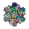 9dwdMC 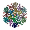 9cwvC 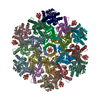 9d6cC 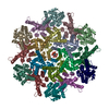 9d6eC 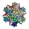 9d88C  9e39C  9p9lC  9p9mC M: atomic model generated by this map C: citing same article ( |
|---|---|
| Similar structure data |


-Links
| EMDB pages | EMDB (EBI/PDBe) / EMDataResource |
|---|---|
| Related items in Molecule of the Month |


-Map
| File | Download / File: emd_47240.map.gz / Format: CCP4 / Size: 158 MB / Type: IMAGE STORED AS FLOATING POINT NUMBER (4 BYTES) | ||||||||||||||||||||||||||||||||||||
|---|---|---|---|---|---|---|---|---|---|---|---|---|---|---|---|---|---|---|---|---|---|---|---|---|---|---|---|---|---|---|---|---|---|---|---|---|---|
| Annotation | anisotropic sharpened map | ||||||||||||||||||||||||||||||||||||
| Projections & slices | Image control
Images are generated by Spider. | ||||||||||||||||||||||||||||||||||||
| Voxel size | X=Y=Z: 0.86 Å | ||||||||||||||||||||||||||||||||||||
| Density |
| ||||||||||||||||||||||||||||||||||||
| Symmetry | Space group: 1 | ||||||||||||||||||||||||||||||||||||
| Details | EMDB XML:
|
 X (Sec.)
 Y (Row.)
 Z (Col.)


-Supplemental data
-Mask #1
| File | emd_47240_msk_1.map | ||||||||||||
|---|---|---|---|---|---|---|---|---|---|---|---|---|---|
| Projections & Slices |
| ||||||||||||
| Density Histograms |


-Additional map: Map without sharpening
| File | emd_47240_additional_1.map | ||||||||||||
|---|---|---|---|---|---|---|---|---|---|---|---|---|---|
| Annotation | Map without sharpening | ||||||||||||
| Projections & Slices |
| ||||||||||||
| Density Histograms |


-Half map: #2
| File | emd_47240_half_map_1.map | ||||||||||||
|---|---|---|---|---|---|---|---|---|---|---|---|---|---|
| Projections & Slices |
| ||||||||||||
| Density Histograms |


-Half map: #1
| File | emd_47240_half_map_2.map | ||||||||||||
|---|---|---|---|---|---|---|---|---|---|---|---|---|---|
| Projections & Slices |
| ||||||||||||
| Density Histograms |


- Sample components
-Entire : Human immunodeficiency virus type 1 (NEW YORK-5 ISOLATE)
| Entire | Name: Human immunodeficiency virus type 1 (NEW YORK-5 ISOLATE) |
|---|---|
| Components |
|
-Supramolecule #1: Human immunodeficiency virus type 1 (NEW YORK-5 ISOLATE)
| Supramolecule | Name: Human immunodeficiency virus type 1 (NEW YORK-5 ISOLATE) type: virus / ID: 1 / Parent: 0 / Macromolecule list: #1 Details: A codon-optimized Gag plasmid with T8I mutation in the SP1 domain (pCMV-Gag-opt) was expressed in HEK293T cells. The enveloped virion like particles were purified by filtration and ...Details: A codon-optimized Gag plasmid with T8I mutation in the SP1 domain (pCMV-Gag-opt) was expressed in HEK293T cells. The enveloped virion like particles were purified by filtration and ultracentrifugation, and directly used as cryo-EM sample. NCBI-ID: 11698 Sci species name: Human immunodeficiency virus type 1 (NEW YORK-5 ISOLATE) Sci species strain: Clone pNL4-3 / Virus type: VIRUS-LIKE PARTICLE / Virus isolate: STRAIN / Virus enveloped: Yes / Virus empty: Yes |
|---|---|
| Host (natural) | Organism:  Homo sapiens (human) Homo sapiens (human) |
| Virus shell | Shell ID: 1 / Name: Gag |
-Macromolecule #1: Gag
| Macromolecule | Name: Gag / type: protein_or_peptide / ID: 1 / Number of copies: 18 / Enantiomer: LEVO |
|---|---|
| Source (natural) | Organism: Human immunodeficiency virus type 1 (NEW YORK-5 ISOLATE) Strain: Clone pNL4-3 |
| Molecular weight | Theoretical: 25.700475 KDa |
| Recombinant expression | Organism: Homo sapiens (human) |
| Sequence | String: QMVHQAISPR TLNAWVKVVE EKAFSPEVIP MFSALSEGAT PQDLNTMLNT VGGHQAAMQM LKETINEEAA EWDRVHPVHA GPIAPGQMR EPRGSDIAGT TSTLQEQIGW MTNNPPIPVG EIYKRWIILG LNKIVRMYSP TSILDIRQGP KEPFRDYVDR F YKTLRAEQ ...String: QMVHQAISPR TLNAWVKVVE EKAFSPEVIP MFSALSEGAT PQDLNTMLNT VGGHQAAMQM LKETINEEAA EWDRVHPVHA GPIAPGQMR EPRGSDIAGT TSTLQEQIGW MTNNPPIPVG EIYKRWIILG LNKIVRMYSP TSILDIRQGP KEPFRDYVDR F YKTLRAEQ ASQEVKNWMT ETLLVQNANP DCKTILKALG PAATLEEMMT ACQGVGGPGH KARVLAEAMS QVIN UniProtKB: Gag polyprotein |
-Macromolecule #2: Lenacapavir
| Macromolecule | Name: Lenacapavir / type: ligand / ID: 2 / Number of copies: 18 / Formula: QNG |
|---|---|
| Molecular weight | Theoretical: 968.282 Da |
-Macromolecule #3: INOSITOL HEXAKISPHOSPHATE
| Macromolecule | Name: INOSITOL HEXAKISPHOSPHATE / type: ligand / ID: 3 / Number of copies: 1 / Formula: IHP |
|---|---|
| Molecular weight | Theoretical: 660.035 Da |
| Chemical component information |  ChemComp-IHP: |
-Experimental details
-Structure determination
| Method | cryo EM |
|---|---|
 Processing Processing | single particle reconstruction |
| Aggregation state | particle |
-Sample preparation
| Concentration | 2 mg/mL | ||||||||||||
|---|---|---|---|---|---|---|---|---|---|---|---|---|---|
| Buffer | pH: 7.4 Component:
Details: This is the final buffer in which the enveloped viral-like particle was resuspended. The Gag-CA-SP1 lattice is inside the viral-like particle and thus not in the direct environment of this buffer. | ||||||||||||
| Vitrification | Cryogen name: ETHANE / Chamber humidity: 100 % / Chamber temperature: 295 K / Instrument: FEI VITROBOT MARK III | ||||||||||||
| Details | A codon-optimized Gag plasmid with T8I mutation in the SP1 domain (pCMV-Gag-opt) was expressed in HEK293T cells. The enveloped virion like particles were purified by filtration and ultracentrifugation, and directly used as cryo-EM sample. |
- Electron microscopy
| Microscope | TFS GLACIOS |
|---|---|
| Image recording | Film or detector model: GATAN K3 BIOQUANTUM (6k x 4k) / Average electron dose: 50.0 e/Å2 |
| Electron beam | Acceleration voltage: 200 kV / Electron source:  FIELD EMISSION GUN FIELD EMISSION GUN |
| Electron optics | C2 aperture diameter: 30.0 µm / Illumination mode: FLOOD BEAM / Imaging mode: BRIGHT FIELD / Cs: 2.7 mm / Nominal defocus max: 2.2 µm / Nominal defocus min: 1.2 µm / Nominal magnification: 45000 |
| Sample stage | Specimen holder model: FEI TITAN KRIOS AUTOGRID HOLDER / Cooling holder cryogen: NITROGEN |

