 Movie
Movie Controller

[English] 日本語
 Yorodumi
- EMDB-46967: Refined plasminogen binding group A streptococcus M-like protein ... -
+ Open data

- Basic information
| Entry |  | |||||||||
|---|---|---|---|---|---|---|---|---|---|---|
| Title | Refined plasminogen binding group A streptococcus M-like protein isolate from AP53 bound to human plasminogen full map | |||||||||
 Map data Map data | Refined PAM isolate from EMD-42429 map. | |||||||||
 Sample Sample |
| |||||||||
 Keywords Keywords | PAM / human plasminogen / M-protein / Group A streptococcus / BLOOD CLOTTING | |||||||||
| Biological species |  Streptococcus pyogenes (bacteria) Streptococcus pyogenes (bacteria) | |||||||||
| Method | single particle reconstruction / cryo EM / Resolution: 3.8 Å | |||||||||
 Authors Authors | Readnour BM / Tjia-Fleck SK / Castellino FJ | |||||||||
| Funding support |  United States, 1 items United States, 1 items
| |||||||||
 Citation Citation | Journal: To Be Published Title: Plasminogen binding group A streptococcus M-like protein from AP53 bound to human plasminogen Authors: Readnour BM / Tjia-Fleck SK / McCann NR / Ayinuola YA / Ploplis VA / Castellino FJ | |||||||||
| History |
|
- Structure visualization
| Supplemental images |
|---|

- Downloads & links
-EMDB archive
| Map data | emd_46967.map.gz | 143.6 MB |  EMDB map data format EMDB map data format | |
|---|---|---|---|---|
| Header (meta data) | emd-46967-v30.xmlemd-46967.xml | 9.9 KB 9.9 KB | Display Display | EMDB header |
| FSC (resolution estimation) | emd_46967_fsc.xml | 16.8 KB | Display | FSC data file |
| Images |  emd_46967.png emd_46967.png | 61 KB | ||
| Filedesc metadata | emd-46967.cif.gz | 4 KB | ||
| Archive directory |  http://ftp.pdbj.org/pub/emdb/structures/EMD-46967ftp://ftp.pdbj.org/pub/emdb/structures/EMD-46967 http://ftp.pdbj.org/pub/emdb/structures/EMD-46967ftp://ftp.pdbj.org/pub/emdb/structures/EMD-46967 | HTTPS FTP |
-Related structure data


-Links
| EMDB pages | EMDB (EBI/PDBe) / EMDataResource |
|---|
-Map
| File | Download / File: emd_46967.map.gz / Format: CCP4 / Size: 178 MB / Type: IMAGE STORED AS FLOATING POINT NUMBER (4 BYTES) | ||||||||||||||||||||||||||||||||||||
|---|---|---|---|---|---|---|---|---|---|---|---|---|---|---|---|---|---|---|---|---|---|---|---|---|---|---|---|---|---|---|---|---|---|---|---|---|---|
| Annotation | Refined PAM isolate from EMD-42429 map. | ||||||||||||||||||||||||||||||||||||
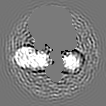
| Projections & slices | Image control
Images are generated by Spider. | ||||||||||||||||||||||||||||||||||||
| Voxel size | X=Y=Z: 3.8 Å | ||||||||||||||||||||||||||||||||||||
| Density |
| ||||||||||||||||||||||||||||||||||||
| Symmetry | Space group: 1 | ||||||||||||||||||||||||||||||||||||
| Details | EMDB XML:
|
 Z (Sec.)
 Y (Row.)
 X (Col.)


-Supplemental data
- Sample components
-Entire : Plasminogen Binding Group A Streptococcus M-Like Protein from AP5...
| Entire | Name: Plasminogen Binding Group A Streptococcus M-Like Protein from AP53 bound to human plasminogen |
|---|---|
| Components |
|
-Supramolecule #1: Plasminogen Binding Group A Streptococcus M-Like Protein from AP5...
| Supramolecule | Name: Plasminogen Binding Group A Streptococcus M-Like Protein from AP53 bound to human plasminogen type: complex / ID: 1 / Parent: 0 / Macromolecule list: #1 / Details: PAM was bound to the surface of a lentivirus |
|---|---|
| Source (natural) | Organism: Streptococcus pyogenes (bacteria) / Strain: AP53 / Location in cell: Cell surface |
| Molecular weight | Theoretical: 41 KDa |
-Experimental details
-Structure determination
| Method | cryo EM |
|---|---|
 Processing Processing | single particle reconstruction |
| Aggregation state | particle |
-Sample preparation
| Buffer | pH: 7.4 / Component - Concentration: 1.0 X / Component - Formula: PBS / Component - Name: PBS |
|---|---|
| Vitrification | Cryogen name: ETHANE / Chamber humidity: 100 % / Chamber temperature: 298 K / Instrument: FEI VITROBOT MARK IV |
| Details | Plasminogen binding Group A streptococcus M like protein of AP53 bound to human plasminogen on the surface of a lentivirus |
- Electron microscopy
| Microscope | TFS KRIOS |
|---|---|
| Image recording | Film or detector model: GATAN K3 (6k x 4k) / Number grids imaged: 2 / Number real images: 4054 / Average electron dose: 55.0 e/Å2 |
| Electron beam | Acceleration voltage: 300 kV / Electron source:  FIELD EMISSION GUN FIELD EMISSION GUN |
| Electron optics | Illumination mode: SPOT SCAN / Imaging mode: BRIGHT FIELD / Cs: 2.7 mm / Nominal defocus max: 34.0 µm / Nominal defocus min: 1.0 µm / Nominal magnification: 109000 |
| Sample stage | Specimen holder model: FEI TITAN KRIOS AUTOGRID HOLDER / Cooling holder cryogen: NITROGEN |
| Experimental equipment |  Model: Titan Krios / Image courtesy: FEI Company |
+Image processing

-Atomic model buiding 1
| Refinement | Space: REAL / Protocol: RIGID BODY FIT |
|---|
