 Movie
Movie Controller

[English] 日本語
 Yorodumi
- EMDB-42429: Plasminogen binding group A streptococcus M-like protein from AP5... -
+ Open data

- Basic information
| Entry |  | ||||||||||||
|---|---|---|---|---|---|---|---|---|---|---|---|---|---|
| Title | Plasminogen binding group A streptococcus M-like protein from AP53 bound to human plasminogen | ||||||||||||
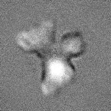
 Map data Map data | Full Map of hPg bound to PAM on a lentivirus | ||||||||||||
 Sample Sample |
| ||||||||||||
 Keywords Keywords | PAM / human plasminogen / M-protein / Group A streptococcus / BLOOD CLOTTING | ||||||||||||
| Function / homology | Plasminogen ligand, VEK-30 / Plasminogen (Pg) ligand in fibrinolytic pathway / : / Streptococcal M proteins D repeats region profile. / : / Streptococcal M proteins C repeat profile. / plasminogen activation / YSIRK Gram-positive signal peptide / Plasminogen-binding group A streptococcal M-like protein PAM Function and homology information Function and homology information | ||||||||||||
| Biological species |  Streptococcus pyogenes (bacteria) Streptococcus pyogenes (bacteria) | ||||||||||||
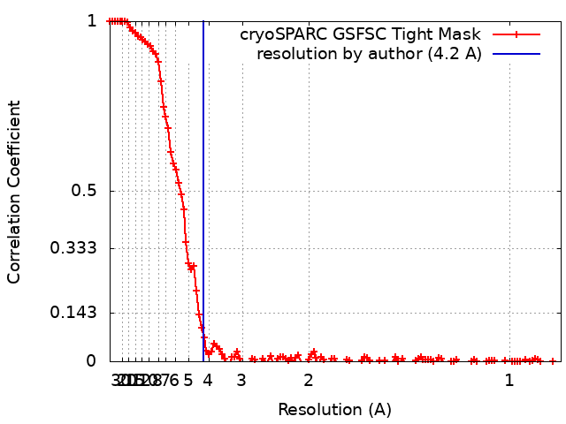
| Method | single particle reconstruction / cryo EM / Resolution: 4.2 Å | ||||||||||||
 Authors Authors | Readnour BM / Tjia-Fleck SK / Castellino FJ / McCann NR | ||||||||||||
| Funding support |  United States, 1 items United States, 1 items
| ||||||||||||
 Citation Citation | Journal: To Be Published Title: Plasminogen binding group A streptococcus M-like protein from AP53 bound to human plasminogen Authors: Readnour BM / Tjia-Fleck SK / McCann NR / Ayinuola YA / Ploplis VA / Castellino FJ | ||||||||||||
| History |
|
- Structure visualization
| Supplemental images |
|---|

- Downloads & links
-EMDB archive
| Map data | emd_42429.map.gz | 166.9 MB | EMDB map data format | |
|---|---|---|---|---|
| Header (meta data) | emd-42429-v30.xmlemd-42429.xml | 15.2 KB 15.2 KB | Display Display | EMDB header |
| FSC (resolution estimation) | emd_42429_fsc.xml | 12 KB | Display | FSC data file |
| Images | 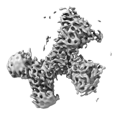 emd_42429.png emd_42429.png | 80.7 KB | ||
| Filedesc metadata | emd-42429.cif.gz | 5 KB | ||
| Others | emd_42429_half_map_1.map.gzemd_42429_half_map_2.map.gz | 165 MB 165 MB | ||
| Archive directory |  http://ftp.pdbj.org/pub/emdb/structures/EMD-42429ftp://ftp.pdbj.org/pub/emdb/structures/EMD-42429 http://ftp.pdbj.org/pub/emdb/structures/EMD-42429ftp://ftp.pdbj.org/pub/emdb/structures/EMD-42429 | HTTPS FTP |
-Related structure data
| Related structure data |  8tvlMC M: atomic model generated by this map C: citing same article ( |
|---|---|
| Similar structure data |

-Links
| EMDB pages | EMDB (EBI/PDBe) / EMDataResource |
|---|
-Map
| File | Download / File: emd_42429.map.gz / Format: CCP4 / Size: 178 MB / Type: IMAGE STORED AS FLOATING POINT NUMBER (4 BYTES) | ||||||||||||||||||||||||||||||||||||
|---|---|---|---|---|---|---|---|---|---|---|---|---|---|---|---|---|---|---|---|---|---|---|---|---|---|---|---|---|---|---|---|---|---|---|---|---|---|
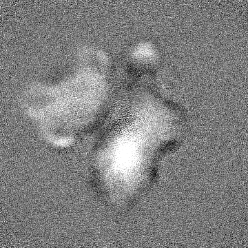
| Annotation | Full Map of hPg bound to PAM on a lentivirus | ||||||||||||||||||||||||||||||||||||
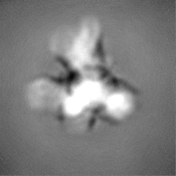
| Projections & slices | Image control
Images are generated by Spider. | ||||||||||||||||||||||||||||||||||||
| Voxel size | X=Y=Z: 0.437 Å | ||||||||||||||||||||||||||||||||||||
| Density |
| ||||||||||||||||||||||||||||||||||||
| Symmetry | Space group: 1 | ||||||||||||||||||||||||||||||||||||
| Details | EMDB XML:
|
 Z (Sec.)
 Y (Row.)
 X (Col.)


-Supplemental data
-Half map: Half Map of hPg bound to PAM on a lentivirus
| File | emd_42429_half_map_1.map | ||||||||||||
|---|---|---|---|---|---|---|---|---|---|---|---|---|---|
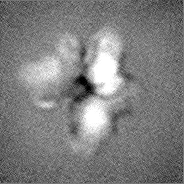
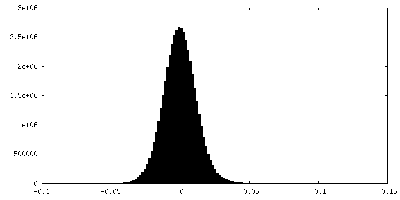
| Annotation | Half Map of hPg bound to PAM on a lentivirus | ||||||||||||
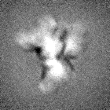
| Projections & Slices |
| ||||||||||||
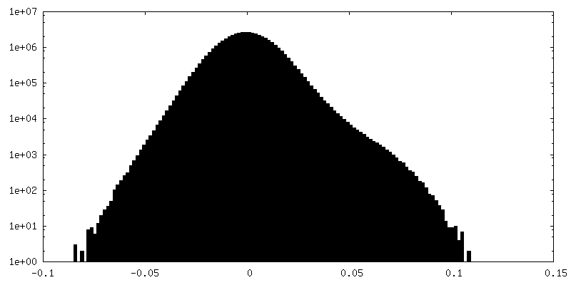
| Density Histograms |


-Half map: Half Map of hPg bound to PAM on a lentivirus
| File | emd_42429_half_map_2.map | ||||||||||||
|---|---|---|---|---|---|---|---|---|---|---|---|---|---|
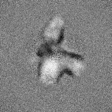
| Annotation | Half Map of hPg bound to PAM on a lentivirus | ||||||||||||
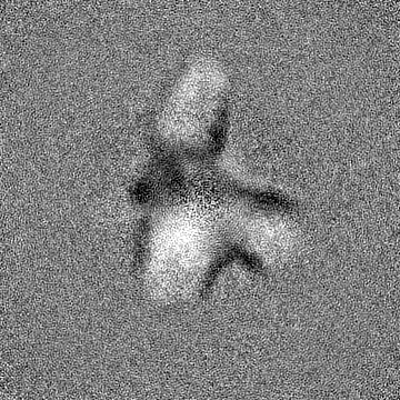
| Projections & Slices |
| ||||||||||||
| Density Histograms |


- Sample components
-Entire : Plasminogen Binding Group A Streptococcus M-Like Protein from AP5...
| Entire | Name: Plasminogen Binding Group A Streptococcus M-Like Protein from AP53 bound to human plasminogen |
|---|---|
| Components |
|
-Supramolecule #1: Plasminogen Binding Group A Streptococcus M-Like Protein from AP5...
| Supramolecule | Name: Plasminogen Binding Group A Streptococcus M-Like Protein from AP53 bound to human plasminogen type: complex / ID: 1 / Parent: 0 / Macromolecule list: all / Details: PAM was bound to the surface of a lentivirus |
|---|---|
| Source (natural) | Organism: Streptococcus pyogenes (bacteria) / Strain: AP53 / Location in cell: Cell surface |
| Molecular weight | Theoretical: 41 KDa |
-Macromolecule #1: Plasminogen binding group A streptococcus M-like protein
| Macromolecule | Name: Plasminogen binding group A streptococcus M-like protein type: protein_or_peptide / ID: 1 / Enantiomer: DEXTRO |
|---|---|
| Source (natural) | Organism: Streptococcus pyogenes (bacteria) / Strain: AP53 |
| Recombinant expression | Organism:  Homo sapiens (human) Homo sapiens (human) |
| Sequence | String: MNRADDARNE VLRGNLVRAE LWYRQIQEND QL KLENKGL KTDLREKEEE LQGLKDDVEK LTADAELQRL KNERHEEAEL ERLKSERHDH DKK EAERKA LEDKLADKQE HLNGALRYIN EKEAEAKEKE AEQKKLKEEK QISDASRQGL RRDL DASRE AKKQVEKDLA ...String: MNRADDARNE VLRGNLVRAE LWYRQIQEND QL KLENKGL KTDLREKEEE LQGLKDDVEK LTADAELQRL KNERHEEAEL ERLKSERHDH DKK EAERKA LEDKLADKQE HLNGALRYIN EKEAEAKEKE AEQKKLKEEK QISDASRQGL RRDL DASRE AKKQVEKDLA NLTAELDKVK EEKQISDASR QGLRRDLDAS REAKKQVEKG LANLT AELD KVKEEKQISD ASRQGLRRDL DASREAKKQV EKALEEANSK LAALEKLNKE LEESKK LTE KEKAELQAKL EAEAKALKEQ LAKQAEELAK LRAEKASDSQ TPDAKPGNKA VPGKGQA PQ AGTKPNQNKA PMKETKRQLP STG |
-Experimental details
-Structure determination
| Method | cryo EM |
|---|---|
 Processing Processing | single particle reconstruction |
| Aggregation state | particle |
-Sample preparation
| Buffer | pH: 7.4 / Component - Formula: PBS |
|---|---|
| Vitrification | Cryogen name: ETHANE / Chamber humidity: 100 % / Chamber temperature: 298 K / Instrument: FEI VITROBOT MARK IV |
| Details | Plasminogen binding Group A streptococcus M like protein of AP53 bound to human plasminogen on the surface of a lentivirus |
- Electron microscopy
| Microscope | TFS KRIOS |
|---|---|
| Image recording | Film or detector model: GATAN K3 (6k x 4k) / Number grids imaged: 2 / Number real images: 4054 / Average electron dose: 55.0 e/Å2 |
| Electron beam | Acceleration voltage: 300 kV / Electron source:  FIELD EMISSION GUN FIELD EMISSION GUN |
| Electron optics | Illumination mode: SPOT SCAN / Imaging mode: BRIGHT FIELD / Cs: 2.7 mm / Nominal defocus max: 34.0 µm / Nominal defocus min: 1.0 µm / Nominal magnification: 109000 |
| Sample stage | Specimen holder model: FEI TITAN KRIOS AUTOGRID HOLDER / Cooling holder cryogen: NITROGEN |
| Experimental equipment |  Model: Titan Krios / Image courtesy: FEI Company |
+Image processing
-Atomic model buiding 1
| Refinement | Space: REAL / Protocol: RIGID BODY FIT |
|---|---|
| Output model | PDB-8tvl: |
