Movie
Movie Controller


+ Open data

- Basic information
| Entry |  | |||||||||
|---|---|---|---|---|---|---|---|---|---|---|
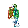
| Title | Structure of URAT1 in complex with lesinurad | |||||||||
 Map data Map data | half map 1 | |||||||||
 Sample Sample |
| |||||||||
 Keywords Keywords | Membrane protein / membrane transporter / TRANSPORT PROTEIN | |||||||||
| Biological species |  Homo sapiens (human) Homo sapiens (human) | |||||||||
| Method | single particle reconstruction / cryo EM / Resolution: 2.74 Å | |||||||||
 Authors Authors | Suo Y / Fedor JG / Lee S-Y | |||||||||
| Funding support |  United States, 1 items United States, 1 items
| |||||||||
 Citation Citation | Journal: Nat Commun / Year: 2025 Title: Molecular basis of the urate transporter URAT1 inhibition by gout drugs. Authors: Yang Suo / Justin G Fedor / Han Zhang / Kalina Tsolova / Xiaoyu Shi / Kedar Sharma / Shweta Kumari / Mario Borgnia / Peng Zhan / Wonpil Im / Seok-Yong Lee /  Abstract: Hyperuricemia is a condition when uric acid, a waste product of purine metabolism, accumulates in the blood. Untreated hyperuricemia can lead to crystal formation of monosodium urate in the joints, ...Hyperuricemia is a condition when uric acid, a waste product of purine metabolism, accumulates in the blood. Untreated hyperuricemia can lead to crystal formation of monosodium urate in the joints, causing a painful inflammatory disease known as gout. These conditions are associated with many other diseases and affect a significant and increasing proportion of the population. The human urate transporter 1 (URAT1) is responsible for the reabsorption of ~90% of uric acid in the kidneys back into the blood, making it a primary target for treating hyperuricemia and gout. Despite decades of research and development, clinically available URAT1 inhibitors have limitations because the molecular basis of URAT1 inhibition by gout drugs remains unknown. Here we present cryo-electron microscopy structures of URAT1 alone and in complex with three clinically relevant inhibitors: benzbromarone, lesinurad, and the recently developed compound TD-3. Together with functional experiments and molecular dynamics simulations, we reveal that these inhibitors bind selectively to URAT1 in inward-open states. Furthermore, we discover differences in the inhibitor-dependent URAT1 conformations as well as interaction networks, which contribute to drug specificity. Our findings illuminate a general theme for URAT1 inhibition, paving the way for the design of next-generation URAT1 inhibitors in the treatment of gout and hyperuricemia. | |||||||||
| History |
|
- Structure visualization
| Supplemental images |
|---|

- Downloads & links
-EMDB archive
| Map data | emd_46950.map.gz | 59.8 MB |  EMDB map data format EMDB map data format | |
|---|---|---|---|---|
| Header (meta data) | emd-46950-v30.xmlemd-46950.xml | 19.5 KB 19.5 KB | Display Display | EMDB header |
| Images |  emd_46950.png emd_46950.png | 81 KB | ||
| Filedesc metadata | emd-46950.cif.gz | 6.6 KB | ||
| Others | emd_46950_half_map_1.map.gzemd_46950_half_map_2.map.gz | 59.4 MB 59.4 MB | ||
| Archive directory |  http://ftp.pdbj.org/pub/emdb/structures/EMD-46950ftp://ftp.pdbj.org/pub/emdb/structures/EMD-46950 http://ftp.pdbj.org/pub/emdb/structures/EMD-46950ftp://ftp.pdbj.org/pub/emdb/structures/EMD-46950 | HTTPS FTP |
-Validation report
| Summary document | emd_46950_validation.pdf.gz | 1.1 MB | Display | EMDB validaton report |
|---|---|---|---|---|
| Full document | emd_46950_full_validation.pdf.gz | 1.1 MB | Display | |
| Data in XML | emd_46950_validation.xml.gz | 12.4 KB | Display | |
| Data in CIF | emd_46950_validation.cif.gz | 14.5 KB | Display | |
| Arichive directory | https://ftp.pdbj.org/pub/emdb/validation_reports/EMD-46950ftp://ftp.pdbj.org/pub/emdb/validation_reports/EMD-46950 | HTTPS FTP |
-Related structure data
| Related structure data |  9dkbMC  9dk9C  9dkaC  9dkcC M: atomic model generated by this map C: citing same article ( |
|---|


-Links
| EMDB pages | EMDB (EBI/PDBe) / EMDataResource |
|---|
-Map
| File | Download / File: emd_46950.map.gz / Format: CCP4 / Size: 64 MB / Type: IMAGE STORED AS FLOATING POINT NUMBER (4 BYTES) | ||||||||||||||||||||||||||||||||||||
|---|---|---|---|---|---|---|---|---|---|---|---|---|---|---|---|---|---|---|---|---|---|---|---|---|---|---|---|---|---|---|---|---|---|---|---|---|---|
| Annotation | half map 1 | ||||||||||||||||||||||||||||||||||||
| Projections & slices | Image control
Images are generated by Spider. | ||||||||||||||||||||||||||||||||||||
| Voxel size | X=Y=Z: 0.8256 Å | ||||||||||||||||||||||||||||||||||||
| Density |
| ||||||||||||||||||||||||||||||||||||
| Symmetry | Space group: 1 | ||||||||||||||||||||||||||||||||||||
| Details | EMDB XML:
|
 Z (Sec.)
 Y (Row.)
 X (Col.)


-Supplemental data
-Half map: half map 1
| File | emd_46950_half_map_1.map | ||||||||||||
|---|---|---|---|---|---|---|---|---|---|---|---|---|---|
| Annotation | half map 1 | ||||||||||||
| Projections & Slices |
| ||||||||||||
| Density Histograms |


-Half map: half map 2
| File | emd_46950_half_map_2.map | ||||||||||||
|---|---|---|---|---|---|---|---|---|---|---|---|---|---|
| Annotation | half map 2 | ||||||||||||
| Projections & Slices |
| ||||||||||||
| Density Histograms |


- Sample components
-Entire : URAT1
| Entire | Name: URAT1 |
|---|---|
| Components |
|
-Supramolecule #1: URAT1
| Supramolecule | Name: URAT1 / type: complex / ID: 1 / Parent: 0 / Macromolecule list: #1 |
|---|---|
| Source (natural) | Organism: Homo sapiens (human) |
| Molecular weight | Theoretical: 55 KDa |
-Macromolecule #1: URAT1
| Macromolecule | Name: URAT1 / type: protein_or_peptide / ID: 1 / Number of copies: 1 / Enantiomer: LEVO |
|---|---|
| Source (natural) | Organism: Homo sapiens (human) |
| Molecular weight | Theoretical: 55.379844 KDa |
| Recombinant expression | Organism: Homo sapiens (human) |
| Sequence | String: MAFSELLDQV GGLGRFQVLQ TVALVVPIMW LCTQSMLENF SAAVPSHRCW VPLLDNSTAQ ASVPGALGPE ALLAVSIPPG PNQGPHQCR RFRQPQWQLL DPNATATNWS EAATEPCVDG WVYDRSTFTS TIVAKWDLVC DSQALKPMAQ SIYLAGILVG A AVCGPASD ...String: MAFSELLDQV GGLGRFQVLQ TVALVVPIMW LCTQSMLENF SAAVPSHRCW VPLLDNSTAQ ASVPGALGPE ALLAVSIPPG PNQGPHQCR RFRQPQWQLL DPNATATNWS EAATEPCVDG WVYDRSTFTS TIVAKWDLVC DSQALKPMAQ SIYLAGILVG A AVCGPASD RFGRRLVLTW SYLQMAVSGT AAAFAPTFPV YCLFRFLVAF AVAGVMMNTG TLVMEWTSAQ ARPLVMTLNS LG FSFGHVL MAAVAYGVRD WALLQLVVSV PFFLCFVYSC WLAESARWLL ITGRLDRGLR ELQRVAAING KRAVGDTLTP QVL LSAMQE ELSVGQAPAS LGTLLRTPGL RLRTCISTLC WFAFGFTFFG LALDLQALGS NIFLLQVLIG VVDIPAKIGT LLLL SRLGR RPTQAASLVL AGLCILANTL VPHEMGALRS ALAVLGLGGL GAAFTCITIY SGELFPTVLR MTAVGLGQMA ARGGA ILGP LVRLLGVHGP WLPLLVYGTV PVLSGLAALL LPET |
-Macromolecule #2: lesinurad
| Macromolecule | Name: lesinurad / type: ligand / ID: 2 / Number of copies: 1 / Formula: A1AIL |
|---|---|
| Molecular weight | Theoretical: 404.281 Da |
-Experimental details
-Structure determination
| Method | cryo EM |
|---|---|
 Processing Processing | single particle reconstruction |
| Aggregation state | particle |
-Sample preparation
| Concentration | 10 mg/mL | ||||||||||||
|---|---|---|---|---|---|---|---|---|---|---|---|---|---|
| Buffer | pH: 8 Component:
| ||||||||||||
| Grid | Model: Quantifoil R1.2/1.3 / Support film - Material: GOLD / Support film - topology: HOLEY / Pretreatment - Type: GLOW DISCHARGE / Pretreatment - Time: 300 sec. / Pretreatment - Atmosphere: AIR / Pretreatment - Pressure: 0.00039000000000000005 kPa | ||||||||||||
| Vitrification | Cryogen name: ETHANE / Chamber humidity: 95 % / Chamber temperature: 280 K / Instrument: LEICA EM GP | ||||||||||||
| Details | Monodisperse sample |
- Electron microscopy
| Microscope | TFS KRIOS |
|---|---|
| Temperature | Max: 70.0 K |
| Specialist optics | Energy filter - Name: GIF Bioquantum / Energy filter - Slit width: 20 eV |
| Image recording | Film or detector model: GATAN K3 (6k x 4k) / Digitization - Dimensions - Width: 5760 pixel / Digitization - Dimensions - Height: 4092 pixel / Number grids imaged: 1 / Number real images: 18880 / Average exposure time: 1.8 sec. / Average electron dose: 50.0 e/Å2 |
| Electron beam | Acceleration voltage: 300 kV / Electron source:  FIELD EMISSION GUN FIELD EMISSION GUN |
| Electron optics | C2 aperture diameter: 100.0 µm / Illumination mode: FLOOD BEAM / Imaging mode: BRIGHT FIELD / Cs: 2.7 mm / Nominal defocus max: 2.0 µm / Nominal defocus min: 1.0 µm / Nominal magnification: 105000 |
| Sample stage | Specimen holder model: FEI TITAN KRIOS AUTOGRID HOLDER / Cooling holder cryogen: NITROGEN |
| Experimental equipment |  Model: Titan Krios / Image courtesy: FEI Company |