 Movie
Movie Controller


+ Open data

- Basic information
| Entry |  | |||||||||
|---|---|---|---|---|---|---|---|---|---|---|
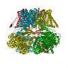
| Title | Ca2+ bound aplysia Slo1 - R196Q | |||||||||
 Map data Map data | ||||||||||
 Sample Sample |
| |||||||||
 Keywords Keywords | Potassium Channel / Voltage sensor / calcium bound / open channel / MEMBRANE PROTEIN | |||||||||
| Function / homology |  Function and homology information Function and homology informationlarge conductance calcium-activated potassium channel activity / monoatomic ion channel complex / modulation of chemical synaptic transmission / postsynaptic membrane / response to xenobiotic stimulus / metal ion binding Similarity search - Function | |||||||||
| Biological species |  | |||||||||
| Method | single particle reconstruction / cryo EM / Resolution: 3.1 Å | |||||||||
 Authors Authors | Contreras GF / Shen R / Latorre R / Perozo E | |||||||||
| Funding support |  United States, 1 items United States, 1 items
| |||||||||
 Citation Citation | Journal: Nat Commun / Year: 2025 Title: Structural basis of voltage-dependent gating in BK channels. Authors: Gustavo F Contreras / Rong Shen / Ramon Latorre / Eduardo Perozo /  Abstract: The allosteric communication between the pore domain, voltage sensors, and Ca binding sites in the calcium- and voltage-activated K channel (BK) underlies its physiological role as the preeminent ...The allosteric communication between the pore domain, voltage sensors, and Ca binding sites in the calcium- and voltage-activated K channel (BK) underlies its physiological role as the preeminent signal integrator in excitable systems. BK displays shallow voltage sensitivity with very fast gating charge kinetics, yet little is known about the molecular underpinnings of this distinctive behavior. Here, we explore the mechanistic basis of coupling between voltage-sensing domains (VSDs) and calcium sensors in Aplysia BK by locking the VSDs in their activated (R196Q and R199Q) and resting (R202Q) states, with or without calcium. Cryo-EM structures of these mutants reveal unique tilts at the S4 C-terminal end, together with large side-chain rotameric excursions of the gating charges. Notably, the VSD resting structure (R202Q) also revealed BK in its elusive, fully closed state, highlighting the reciprocal relation between calcium and voltage sensors. These structures provide a plausible path where voltage and Ca binding couple energetically and define the conformation of the pore domain and, thus, BK's full functional range. | |||||||||
| History |
|
- Structure visualization
| Supplemental images |
|---|

- Downloads & links
-EMDB archive
| Map data | emd_46901.map.gz | 45.5 MB | EMDB map data format | |
|---|---|---|---|---|
| Header (meta data) | emd-46901-v30.xmlemd-46901.xml | 16.6 KB 16.6 KB | Display Display | EMDB header |
| FSC (resolution estimation) | emd_46901_fsc.xml | 11.7 KB | Display | FSC data file |
| Images |  emd_46901.png emd_46901.png | 151.7 KB | ||
| Filedesc metadata | emd-46901.cif.gz | 6.3 KB | ||
| Others | emd_46901_half_map_1.map.gzemd_46901_half_map_2.map.gz | 45.7 MB 45.7 MB | ||
| Archive directory |  http://ftp.pdbj.org/pub/emdb/structures/EMD-46901ftp://ftp.pdbj.org/pub/emdb/structures/EMD-46901 http://ftp.pdbj.org/pub/emdb/structures/EMD-46901ftp://ftp.pdbj.org/pub/emdb/structures/EMD-46901 | HTTPS FTP |
-Related structure data
| Related structure data |  9di8MC  9dicC  9ditC  9djvC  9dkfC  9dklC  9dknC M: atomic model generated by this map C: citing same article ( |
|---|---|
| Similar structure data |


-Links
| EMDB pages | EMDB (EBI/PDBe) / EMDataResource |
|---|---|
| Related items in Molecule of the Month |


-Map
| File | Download / File: emd_46901.map.gz / Format: CCP4 / Size: 64 MB / Type: IMAGE STORED AS FLOATING POINT NUMBER (4 BYTES) | ||||||||||||||||||||||||||||||||||||
|---|---|---|---|---|---|---|---|---|---|---|---|---|---|---|---|---|---|---|---|---|---|---|---|---|---|---|---|---|---|---|---|---|---|---|---|---|---|
| Projections & slices | Image control
Images are generated by Spider. | ||||||||||||||||||||||||||||||||||||
| Voxel size | X=Y=Z: 1.063 Å | ||||||||||||||||||||||||||||||||||||
| Density |
| ||||||||||||||||||||||||||||||||||||
| Symmetry | Space group: 1 | ||||||||||||||||||||||||||||||||||||
| Details | EMDB XML:
|
 Z (Sec.)
 Y (Row.)
 X (Col.)


-Supplemental data
-Half map: #2
| File | emd_46901_half_map_1.map | ||||||||||||
|---|---|---|---|---|---|---|---|---|---|---|---|---|---|
| Projections & Slices |
| ||||||||||||
| Density Histograms |


-Half map: #1
| File | emd_46901_half_map_2.map | ||||||||||||
|---|---|---|---|---|---|---|---|---|---|---|---|---|---|
| Projections & Slices |
| ||||||||||||
| Density Histograms |


- Sample components
-Entire : homotetramer large-conductance calcium- and voltage-activated K+ ...
| Entire | Name: homotetramer large-conductance calcium- and voltage-activated K+ channel |
|---|---|
| Components |
|
-Supramolecule #1: homotetramer large-conductance calcium- and voltage-activated K+ ...
| Supramolecule | Name: homotetramer large-conductance calcium- and voltage-activated K+ channel type: complex / ID: 1 / Parent: 0 / Macromolecule list: #1 |
|---|---|
| Source (natural) | Organism: |
-Macromolecule #1: BK channel
| Macromolecule | Name: BK channel / type: protein_or_peptide / ID: 1 / Number of copies: 4 / Enantiomer: LEVO |
|---|---|
| Source (natural) | Organism: |
| Molecular weight | Theoretical: 119.684297 KDa |
| Recombinant expression | Organism:   Spodoptera frugiperda (fall armyworm) Spodoptera frugiperda (fall armyworm) |
| Sequence | String: MASSSSTSCE PGDRQWYSFL ASSLVTFGSG LVVIIIYRIV LWLCCRKKKC IQVSNPVPTA RTTSLDQKSF MKNSDPEIGW MTEAKDWAG ELISGQTTTG RILVGLVFLL SIASLIIYFI DASTNTSVET CLPWSSSTTQ QVDLAFNVFF MIYFFIRFVA A NDKLWFWV ...String: MASSSSTSCE PGDRQWYSFL ASSLVTFGSG LVVIIIYRIV LWLCCRKKKC IQVSNPVPTA RTTSLDQKSF MKNSDPEIGW MTEAKDWAG ELISGQTTTG RILVGLVFLL SIASLIIYFI DASTNTSVET CLPWSSSTTQ QVDLAFNVFF MIYFFIRFVA A NDKLWFWV ELFSFVDYFT IPPSFVAIYL DRNWLGLQFL RALRLMSIPD ILTYLNVLKT STLIRLVQLV VSFVSLWLTA AG FLHLLEN SGDPFFDFGN AQHLTYWECL YFLMVTMSTV GFGDIFATTV LGRTFVVIFI MIFIGLFASF IPEIAEILGK RQK YGGSYK KERGKRHVVV CGYITFDSVS NFLKDFLHKD REDVDVEIVF LHKGLPGLEL EGLLKRHFTQ VEYFWGSVMD ANDL ERVKI QEADACLVLA NKYCQDPDQE DAANIMRVIS IKNYHSDIKV IVQLLQYHNK AYLLNIPSWD WKRGDDAVCV AELKL GFIA QSCLAPGFST LMANLFTMRS YKPTPEMSQW QTDYMRGTGM EMYTEYLSSA FNALTFPEAA ELCFSKLKLL LLAIEV RQE DTRESTLAIN PGPKVKIENA TQGFFIAESA EEVKRAFYYC KNCHANVSDV RQIKKCKCRP LAMFKKGAAA VLALQRT PG LAVEPDGEAN DKDKSRGTST SKAVTSFPEK RKPQSRRKPS TTLKSKSPSE DSVPPPPPPV DEPRKFDSTG MFHWCPDR P LNDCLQDRSQ ASASGLRNHV VVCLFADAAS PLIGLRNLVM PLRASNFHYH ELKPTIIVGN LDYLHREWKT LQNFPKLSI LPGSPLNRAN LRAVNINLCD MCVIVSAKDR NMEDPNLVDK EAILCSLNIK AMTFDDTMGL IQSSNFVPGG FSPLHENKRS QAGANVPLI TELANDSNVQ FLDQDDDDDP DTELYMTQPF ACGTAFAVSV LDSLMSTSYF NDNALTLIRT LITGGATPEL E QILAEGAG MRGGYCSPAV LANRDRCRVA QISLFDGPLA QFGQGGHYGE LFVYALRHFG ILCIGLYRFR DTNESVRSPS SK RYVITNP PEDFPLLPTD QVYVLTYK UniProtKB: BK channel |
-Macromolecule #2: POTASSIUM ION
| Macromolecule | Name: POTASSIUM ION / type: ligand / ID: 2 / Number of copies: 5 / Formula: K |
|---|---|
| Molecular weight | Theoretical: 39.098 Da |
-Macromolecule #3: MAGNESIUM ION
| Macromolecule | Name: MAGNESIUM ION / type: ligand / ID: 3 / Number of copies: 4 / Formula: MG |
|---|---|
| Molecular weight | Theoretical: 24.305 Da |
-Macromolecule #4: CALCIUM ION
| Macromolecule | Name: CALCIUM ION / type: ligand / ID: 4 / Number of copies: 7 / Formula: CA |
|---|---|
| Molecular weight | Theoretical: 40.078 Da |
-Experimental details
-Structure determination
| Method | cryo EM |
|---|---|
 Processing Processing | single particle reconstruction |
| Aggregation state | particle |
-Sample preparation
| Buffer | pH: 8 |
|---|---|
| Vitrification | Cryogen name: ETHANE |
- Electron microscopy
| Microscope | FEI TITAN KRIOS |
|---|---|
| Image recording | Film or detector model: GATAN K3 BIOQUANTUM (6k x 4k) / Average electron dose: 65.0 e/Å2 |
| Electron beam | Acceleration voltage: 300 kV / Electron source:  FIELD EMISSION GUN FIELD EMISSION GUN |
| Electron optics | Illumination mode: OTHER / Imaging mode: DIFFRACTION / Nominal defocus max: 2.2 µm / Nominal defocus min: 0.5 µm |
| Experimental equipment |  Model: Titan Krios / Image courtesy: FEI Company |
+Image processing

-Atomic model buiding 1
| Refinement | Protocol: FLEXIBLE FIT |
|---|---|
| Output model | PDB-9di8: |
