 Movie
Movie Controller


+ Open data

- Basic information
| Entry |  | |||||||||||||||
|---|---|---|---|---|---|---|---|---|---|---|---|---|---|---|---|---|
| Title | Resting state 1 of the GluA2-gamma2 complex | |||||||||||||||
 Map data Map data | Resting state 1 LBD-TMD locally sharpened map | |||||||||||||||
 Sample Sample |
| |||||||||||||||
 Keywords Keywords | ligand-gated ion channel / ionotropic glutamate receptor / ampa receptor / ion channel / TRANSPORT PROTEIN | |||||||||||||||
| Function / homology |  Function and homology information Function and homology informationPresynaptic depolarization and calcium channel opening / LGI-ADAM interactions / Trafficking of AMPA receptors / eye blink reflex / positive regulation of protein localization to basolateral plasma membrane / cerebellar mossy fiber / postsynaptic neurotransmitter receptor diffusion trapping / regulation of AMPA receptor activity / channel regulator activity / membrane hyperpolarization ...Presynaptic depolarization and calcium channel opening / LGI-ADAM interactions / Trafficking of AMPA receptors / eye blink reflex / positive regulation of protein localization to basolateral plasma membrane / cerebellar mossy fiber / postsynaptic neurotransmitter receptor diffusion trapping / regulation of AMPA receptor activity / channel regulator activity / membrane hyperpolarization / nervous system process / protein targeting to membrane / voltage-gated calcium channel complex / spine synapse / dendritic spine neck / dendritic spine cytoplasm / dendritic spine head / cellular response to amine stimulus / Activation of AMPA receptors / neurotransmitter receptor localization to postsynaptic specialization membrane / ligand-gated monoatomic cation channel activity / perisynaptic space / neuromuscular junction development / Trafficking of GluR2-containing AMPA receptors / response to lithium ion / AMPA glutamate receptor activity / AMPA glutamate receptor clustering / transmission of nerve impulse / kainate selective glutamate receptor activity / immunoglobulin binding / AMPA glutamate receptor complex / regulation of receptor recycling / extracellularly glutamate-gated ion channel activity / cellular response to glycine / ionotropic glutamate receptor complex / asymmetric synapse / Unblocking of NMDA receptors, glutamate binding and activation / glutamate receptor binding / membrane depolarization / positive regulation of synaptic transmission / conditioned place preference / regulation of postsynaptic membrane neurotransmitter receptor levels / voltage-gated calcium channel activity / response to fungicide / regulation of synaptic transmission, glutamatergic / extracellular ligand-gated monoatomic ion channel activity / cytoskeletal protein binding / glutamate-gated receptor activity / cellular response to brain-derived neurotrophic factor stimulus / regulation of long-term synaptic depression / positive regulation of synaptic transmission, glutamatergic / somatodendritic compartment / glutamate-gated calcium ion channel activity / presynaptic active zone membrane / excitatory synapse / ionotropic glutamate receptor signaling pathway / ionotropic glutamate receptor binding / dendrite cytoplasm / dendrite membrane / ligand-gated monoatomic ion channel activity involved in regulation of presynaptic membrane potential / positive regulation of excitatory postsynaptic potential / hippocampal mossy fiber to CA3 synapse / dendritic shaft / SNARE binding / synaptic membrane / PDZ domain binding / protein tetramerization / establishment of protein localization / synaptic transmission, glutamatergic / transmitter-gated monoatomic ion channel activity involved in regulation of postsynaptic membrane potential / regulation of membrane potential / cerebral cortex development / response to calcium ion / receptor internalization / postsynaptic density membrane / modulation of chemical synaptic transmission / Schaffer collateral - CA1 synapse / terminal bouton / synaptic vesicle / long-term synaptic potentiation / amyloid-beta binding / synaptic vesicle membrane / growth cone / presynapse / signaling receptor activity / presynaptic membrane / scaffold protein binding / dendritic spine / chemical synaptic transmission / perikaryon / postsynaptic membrane / neuron projection / postsynaptic density / external side of plasma membrane / axon / neuronal cell body / synapse / dendrite / protein kinase binding / protein-containing complex binding Similarity search - Function | |||||||||||||||
| Biological species |  | |||||||||||||||
| Method | single particle reconstruction / cryo EM / Resolution: 4.18 Å | |||||||||||||||
 Authors Authors | Kumar Mondal A / Carrillo E / Jayaraman V / Twomey EC | |||||||||||||||
| Funding support |  United States, 4 items United States, 4 items
| |||||||||||||||
 Citation Citation | Journal: Nature / Year: 2025 Title: Glutamate gating of AMPA-subtype iGluRs at physiological temperatures. Authors: Anish Kumar Mondal / Elisa Carrillo / Vasanthi Jayaraman / Edward C Twomey / Abstract: Ionotropic glutamate receptors (iGluRs) are tetrameric ligand-gated ion channels that mediate most excitatory neurotransmission. iGluRs are gated by glutamate, where on glutamate binding, they open ...Ionotropic glutamate receptors (iGluRs) are tetrameric ligand-gated ion channels that mediate most excitatory neurotransmission. iGluRs are gated by glutamate, where on glutamate binding, they open their ion channels to enable cation influx into postsynaptic neurons, initiating signal transduction. The structural mechanics of how glutamate gating occurs in full-length iGluRs is not well understood. Here, using the α-amino-3-hydroxy-5-methyl-4-isoxazolepropionic acid subtype iGluR (AMPAR), we identify the glutamate-gating mechanism. AMPAR activation by glutamate is augmented at physiological temperatures. By preparing AMPARs for cryogenic-electron microscopy at these temperatures, we captured the glutamate-gating mechanism. Activation by glutamate initiates ion channel opening that involves all ion channel helices hinging away from the pore axis in a motif that is conserved across all iGluRs. Desensitization occurs when the local dimer pairs decouple and enables closure of the ion channel below through restoring the channel hinges and refolding the channel gate. Our findings define how glutamate gates iGluRs, provide foundations for therapeutic design and demonstrate how physiological temperatures can alter iGluR function. | |||||||||||||||
| History |
|
- Structure visualization
| Supplemental images |
|---|

- Downloads & links
-EMDB archive
| Map data | emd_46872.map.gz | 5.9 MB | EMDB map data format | |
|---|---|---|---|---|
| Header (meta data) | emd-46872-v30.xmlemd-46872.xml | 18.6 KB 18.6 KB | Display Display | EMDB header |
| Images |  emd_46872.png emd_46872.png | 41.8 KB | ||
| Filedesc metadata | emd-46872.cif.gz | 6.2 KB | ||
| Others | emd_46872_half_map_1.map.gzemd_46872_half_map_2.map.gz | 300.7 MB 300.7 MB | ||
| Archive directory |  http://ftp.pdbj.org/pub/emdb/structures/EMD-46872ftp://ftp.pdbj.org/pub/emdb/structures/EMD-46872 http://ftp.pdbj.org/pub/emdb/structures/EMD-46872ftp://ftp.pdbj.org/pub/emdb/structures/EMD-46872 | HTTPS FTP |
-Related structure data
| Related structure data | 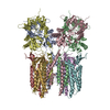 9dhpMC 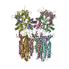 9dhqC 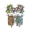 9dhrC 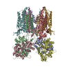 9dhsC 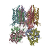 9dhtC 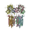 9mrkC 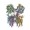 9mrlC 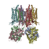 9mrmC 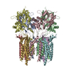 9mrnC M: atomic model generated by this map C: citing same article ( |
|---|---|
| Similar structure data |


-Links
| EMDB pages | EMDB (EBI/PDBe) / EMDataResource |
|---|---|
| Related items in Molecule of the Month |


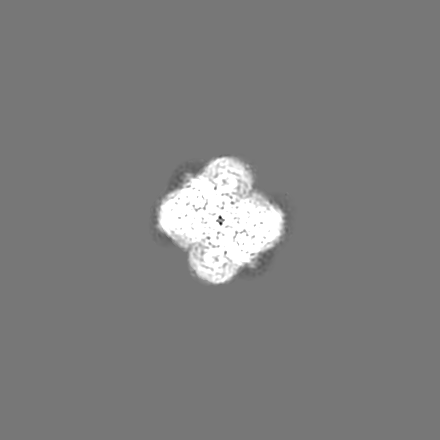
-Map
| File | Download / File: emd_46872.map.gz / Format: CCP4 / Size: 325 MB / Type: IMAGE STORED AS FLOATING POINT NUMBER (4 BYTES) | ||||||||||||||||||||||||||||||||||||
|---|---|---|---|---|---|---|---|---|---|---|---|---|---|---|---|---|---|---|---|---|---|---|---|---|---|---|---|---|---|---|---|---|---|---|---|---|---|
| Annotation | Resting state 1 LBD-TMD locally sharpened map | ||||||||||||||||||||||||||||||||||||
| Projections & slices | Image control
Images are generated by Spider. | ||||||||||||||||||||||||||||||||||||
| Voxel size | X=Y=Z: 0.97 Å | ||||||||||||||||||||||||||||||||||||
| Density |
| ||||||||||||||||||||||||||||||||||||
| Symmetry | Space group: 1 | ||||||||||||||||||||||||||||||||||||
| Details | EMDB XML:
|
 Z (Sec.)
 Y (Row.)
 X (Col.)


-Supplemental data
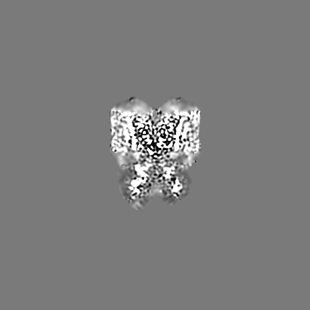
-Half map: Resting state 1 LBD-TMD half map
| File | emd_46872_half_map_1.map | ||||||||||||
|---|---|---|---|---|---|---|---|---|---|---|---|---|---|
| Annotation | Resting state 1 LBD-TMD half map | ||||||||||||
| Projections & Slices |
| ||||||||||||
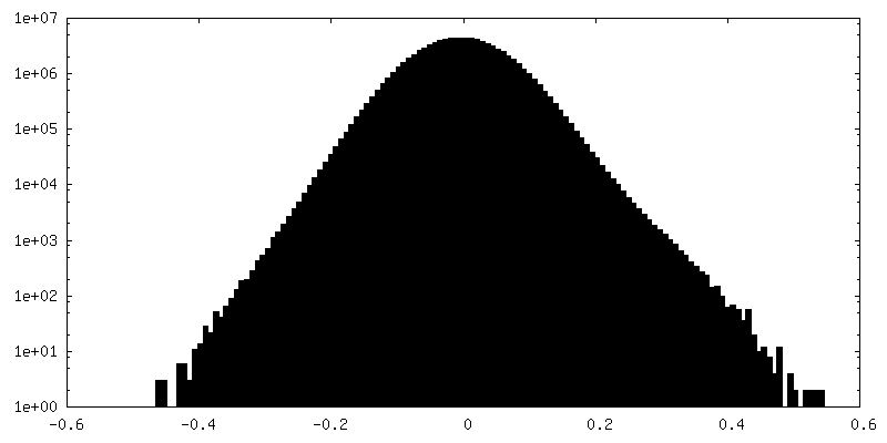
| Density Histograms |


-Half map: Resting state 1 LBD-TMD half map
| File | emd_46872_half_map_2.map | ||||||||||||
|---|---|---|---|---|---|---|---|---|---|---|---|---|---|
| Annotation | Resting state 1 LBD-TMD half map | ||||||||||||
| Projections & Slices |
| ||||||||||||
| Density Histograms |


- Sample components
-Entire : GluA2-TARPgamma2 Complex
| Entire | Name: GluA2-TARPgamma2 Complex |
|---|---|
| Components |
|
-Supramolecule #1: GluA2-TARPgamma2 Complex
| Supramolecule | Name: GluA2-TARPgamma2 Complex / type: complex / ID: 1 / Parent: 0 / Macromolecule list: all |
|---|---|
| Source (natural) | Organism: |
-Macromolecule #1: Isoform Flip of Glutamate receptor 2
| Macromolecule | Name: Isoform Flip of Glutamate receptor 2 / type: protein_or_peptide / ID: 1 / Number of copies: 4 / Enantiomer: LEVO |
|---|---|
| Source (natural) | Organism: |
| Molecular weight | Theoretical: 47.938141 KDa |
| Recombinant expression | Organism:  Homo sapiens (human) Homo sapiens (human) |
| Sequence | String: EQKTVVVTTI LESPYVMMKK NHEMLEGNER YEGYCVDLAA EIAKHCGFKY KLTIVGDGKY GARDADTKIW NGMVGELVYG KADIAIAPL TITLVREEVI DFSKPFMSLG ISIMIKKPQK SKPGVFSFLD PLAYEIWMCI VFAYIGVSVV LFLVSRFSPY E WHTEEFED ...String: EQKTVVVTTI LESPYVMMKK NHEMLEGNER YEGYCVDLAA EIAKHCGFKY KLTIVGDGKY GARDADTKIW NGMVGELVYG KADIAIAPL TITLVREEVI DFSKPFMSLG ISIMIKKPQK SKPGVFSFLD PLAYEIWMCI VFAYIGVSVV LFLVSRFSPY E WHTEEFED GRETQSSEST NEFGIFNSLW FSLGAFMQQG CDISPRSLSG RIVGGVWWFF TLIIISSYTA NLAAFLTVER MV SPIESAE DLSKQTEIAY GTLDSGSTKE FFRRSKIAVF DKMWTYMRSA EPSVFVRTTA EGVARVRKSK GKYAYLLEST MNE YIEQRK PCDTMKVGGN LDSKGYGIAT PKGSSLGTPV NLAVLKLSEQ GVLDKLKNKW WYDKGECGAK DSGSKEKTSA LSLS NVAGV FYILVGGLGL AMLVALIEFC YKSRA UniProtKB: Glutamate receptor 2 |
-Macromolecule #2: Voltage-dependent calcium channel gamma-2 subunit
| Macromolecule | Name: Voltage-dependent calcium channel gamma-2 subunit / type: protein_or_peptide / ID: 2 / Number of copies: 4 / Enantiomer: LEVO |
|---|---|
| Source (natural) | Organism: |
| Molecular weight | Theoretical: 22.633041 KDa |
| Recombinant expression | Organism: Homo sapiens (human) |
| Sequence | String: RGVQMLLTTV GAFAAFSLMT IAVGTDYWLY SRGVCKTKSV SENETSKKNE EVMTHSGLWR TCCLEGNFKG LCKQIDHFPE DADYEADTA EYFLRAVRAS SIFPILSVIL LFMGGLCIAA SEFYKTRHNI ILSAGIFFVS AGLSNIIGII VYISANAGDP S KSDSKKNS ...String: RGVQMLLTTV GAFAAFSLMT IAVGTDYWLY SRGVCKTKSV SENETSKKNE EVMTHSGLWR TCCLEGNFKG LCKQIDHFPE DADYEADTA EYFLRAVRAS SIFPILSVIL LFMGGLCIAA SEFYKTRHNI ILSAGIFFVS AGLSNIIGII VYISANAGDP S KSDSKKNS YSYGWSFYFG ALSFIIAEMV GVLAVHMFID RHKQLTG UniProtKB: Voltage-dependent calcium channel gamma-2 subunit |
-Experimental details
-Structure determination
| Method | cryo EM |
|---|---|
 Processing Processing | single particle reconstruction |
| Aggregation state | particle |
-Sample preparation
| Concentration | 4 mg/mL |
|---|---|
| Buffer | pH: 8 |
| Vitrification | Cryogen name: ETHANE |
- Electron microscopy
| Microscope | TFS KRIOS |
|---|---|
| Image recording | Film or detector model: FEI FALCON IV (4k x 4k) / Average electron dose: 40.0 e/Å2 |
| Electron beam | Acceleration voltage: 300 kV / Electron source:  FIELD EMISSION GUN FIELD EMISSION GUN |
| Electron optics | Illumination mode: FLOOD BEAM / Imaging mode: BRIGHT FIELD / Nominal defocus max: 2.5 µm / Nominal defocus min: 1.0 µm |
| Experimental equipment |  Model: Titan Krios / Image courtesy: FEI Company |
