 Movie
Movie Controller


+ Open data

- Basic information
| Entry |  | |||||||||
|---|---|---|---|---|---|---|---|---|---|---|
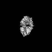
| Title | Mycobacterium tuberculosis UvrD1: DNA-bound dimer. | |||||||||
 Map data Map data | sharpened map | |||||||||
 Sample Sample |
| |||||||||
 Keywords Keywords | DNA Helicase / DNA Translocase / ATPase / DNA BINDING PROTEIN / DNA BINDING PROTEIN-DNA complex | |||||||||
| Function / homology |  Function and homology information Function and homology informationnegative regulation of strand invasion / DNA helicase complex / UV protection / recombinational repair / dATP binding / DNA 3'-5' helicase / 3'-5' DNA helicase activity / ATP-dependent activity, acting on DNA / peptidoglycan-based cell wall / double-strand break repair ...negative regulation of strand invasion / DNA helicase complex / UV protection / recombinational repair / dATP binding / DNA 3'-5' helicase / 3'-5' DNA helicase activity / ATP-dependent activity, acting on DNA / peptidoglycan-based cell wall / double-strand break repair / DNA replication / magnesium ion binding / ATP hydrolysis activity / DNA binding / ATP binding / plasma membrane / cytosol Similarity search - Function | |||||||||
| Biological species |   Mycobacterium tuberculosis (bacteria) Mycobacterium tuberculosis (bacteria) | |||||||||
| Method | single particle reconstruction / cryo EM / Resolution: 7.0 Å | |||||||||
 Authors Authors | Chadda A / Galburt EA | |||||||||
| Funding support |  United States, 1 items United States, 1 items
| |||||||||
 Citation Citation | Journal: Proc Natl Acad Sci U S A / Year: 2025 Title: Structural basis for dimerization and activation of UvrD-family helicases. Authors: Ankita Chadda / Binh Nguyen / Timothy M Lohman / Eric A Galburt / Abstract: UvrD-family helicases are superfamily 1A motor proteins that function during DNA replication, recombination, repair, and transcription. UvrD family monomers translocate along single-stranded (ss) DNA ...UvrD-family helicases are superfamily 1A motor proteins that function during DNA replication, recombination, repair, and transcription. UvrD family monomers translocate along single-stranded (ss) DNA but need to be activated by dimerization to unwind DNA in the absence of force or accessory factors. However, prior structural studies have only revealed monomeric complexes. Here, we report the first structures of a dimeric UvrD-family helicase, UvrD1, both free and bound to a DNA junction. In each structure, the dimer interface occurs between the 2B subdomains of each subunit. The apo UvrD1 dimer is observed in symmetric compact and extended forms indicating substantial flexibility. This symmetry is broken in the DNA-bound dimer complex with leading and trailing subunits adopting distinct conformations. Biochemical experiments reveal that the UvrD dimer shares the same 2B-2B interface. In contrast to the dimeric structures, an inactive, autoinhibited UvrD1 DNA-bound monomer structure reveals 2B subdomain-DNA contacts that are likely inhibitory. The major reorientation of the 2B subdomains that occurs upon UvrD1 dimerization prevents these duplex DNA interactions, thus relieving the autoinhibition. These structures reveal that the 2B subdomain serves a major regulatory role rather than participating directly in DNA unwinding. | |||||||||
| History |
|
- Structure visualization
| Supplemental images |
|---|

- Downloads & links
-EMDB archive
| Map data | emd_46797.map.gz | 28.7 MB | EMDB map data format | |
|---|---|---|---|---|
| Header (meta data) | emd-46797-v30.xmlemd-46797.xml | 20.4 KB 20.4 KB | Display Display | EMDB header |
| FSC (resolution estimation) | emd_46797_fsc.xml | 9.2 KB | Display | FSC data file |
| Images |  emd_46797.png emd_46797.png | 84.4 KB | ||
| Filedesc metadata | emd-46797.cif.gz | 6.7 KB | ||
| Others | emd_46797_additional_1.map.gzemd_46797_half_map_1.map.gzemd_46797_half_map_2.map.gz | 15.1 MB 28.4 MB 28.4 MB | ||
| Archive directory |  http://ftp.pdbj.org/pub/emdb/structures/EMD-46797ftp://ftp.pdbj.org/pub/emdb/structures/EMD-46797 http://ftp.pdbj.org/pub/emdb/structures/EMD-46797ftp://ftp.pdbj.org/pub/emdb/structures/EMD-46797 | HTTPS FTP |
-Related structure data
| Related structure data |  9desMC  9dciC  9dgyC M: atomic model generated by this map C: citing same article ( |
|---|---|
| Similar structure data |


-Links
| EMDB pages | EMDB (EBI/PDBe) / EMDataResource |
|---|---|
| Related items in Molecule of the Month |


-Map
| File | Download / File: emd_46797.map.gz / Format: CCP4 / Size: 30.5 MB / Type: IMAGE STORED AS FLOATING POINT NUMBER (4 BYTES) | ||||||||||||||||||||||||||||||||||||
|---|---|---|---|---|---|---|---|---|---|---|---|---|---|---|---|---|---|---|---|---|---|---|---|---|---|---|---|---|---|---|---|---|---|---|---|---|---|
| Annotation | sharpened map | ||||||||||||||||||||||||||||||||||||
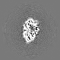
| Projections & slices | Image control
Images are generated by Spider. | ||||||||||||||||||||||||||||||||||||
| Voxel size | X=Y=Z: 1.624 Å | ||||||||||||||||||||||||||||||||||||
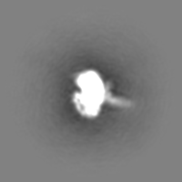
| Density |
| ||||||||||||||||||||||||||||||||||||
| Symmetry | Space group: 1 | ||||||||||||||||||||||||||||||||||||
| Details | EMDB XML:
|
 Z (Sec.)
 Y (Row.)
 X (Col.)


-Supplemental data
-Additional map: unsharpened map
| File | emd_46797_additional_1.map | ||||||||||||
|---|---|---|---|---|---|---|---|---|---|---|---|---|---|
| Annotation | unsharpened map | ||||||||||||
| Projections & Slices |
| ||||||||||||
| Density Histograms |


-Half map: half map B
| File | emd_46797_half_map_1.map | ||||||||||||
|---|---|---|---|---|---|---|---|---|---|---|---|---|---|
| Annotation | half map B | ||||||||||||
| Projections & Slices |
| ||||||||||||
| Density Histograms |


-Half map: half map A
| File | emd_46797_half_map_2.map | ||||||||||||
|---|---|---|---|---|---|---|---|---|---|---|---|---|---|
| Annotation | half map A | ||||||||||||
| Projections & Slices |
| ||||||||||||
| Density Histograms |


- Sample components
-Entire : UvrD1 dimer bound to dsDNA-ssDNA junction
| Entire | Name: UvrD1 dimer bound to dsDNA-ssDNA junction |
|---|---|
| Components |
|
-Supramolecule #1: UvrD1 dimer bound to dsDNA-ssDNA junction
| Supramolecule | Name: UvrD1 dimer bound to dsDNA-ssDNA junction / type: complex / ID: 1 / Parent: 0 / Macromolecule list: all |
|---|---|
| Source (natural) | Organism: Mycobacterium tuberculosis (bacteria) |
| Molecular weight | Theoretical: 170 KDa |
-Macromolecule #1: ATP-dependent DNA helicase UvrD1
| Macromolecule | Name: ATP-dependent DNA helicase UvrD1 / type: protein_or_peptide / ID: 1 / Number of copies: 2 / Enantiomer: LEVO / EC number: DNA 3'-5' helicase |
|---|---|
| Source (natural) | Organism: Mycobacterium tuberculosis (bacteria) |
| Molecular weight | Theoretical: 85.154898 KDa |
| Recombinant expression | Organism: |
| Sequence | String: MSVHATDAKP PGPSPADQLL DGLNPQQRQA VVHEGSPLLI VAGAGSGKTA VLTRRIAYLM AARGVGVGQI LAITFTNKAA AEMRERVVG LVGEKARYMW VSTFHSTCVR ILRNQAALIE GLNSNFSIYD ADDSRRLLQM VGRDLGLDIK RYSPRLLANA I SNLKNELI ...String: MSVHATDAKP PGPSPADQLL DGLNPQQRQA VVHEGSPLLI VAGAGSGKTA VLTRRIAYLM AARGVGVGQI LAITFTNKAA AEMRERVVG LVGEKARYMW VSTFHSTCVR ILRNQAALIE GLNSNFSIYD ADDSRRLLQM VGRDLGLDIK RYSPRLLANA I SNLKNELI DPHQALAGLT EDSDDLARAV ASVYDEYQRR LRAANALDFD DLIGETVAVL QAFPQIAQYY RRRFRHVLVD EY QDTNHAQ YVLVRELVGR DSNDGIPPGE LCVVGDADQS IYAFRGATIR NIEDFERDYP DTRTILLEQN YRSTQNILSA ANS VIARNA GRREKRLWTD AGAGELIVGY VADNEHDEAR FVAEEIDALA EGSEITYNDV AVFYRTNNSS RSLEEVLIRA GIPY KVVGG VRFYERKEIR DIVAYLRVLD NPGDAVSLRR ILNTPRRGIG DRAEACVAVY AENTGVGFGD ALVAAAQGKV PMLNT RAEK AIAGFVEMFD ELRGRLDDDL GELVEAVLER TGYRRELEAS TDPQELARLD NLNELVSVAH EFSTDRENAA ALGPDD EDV PDTGVLADFL ERVSLVADAD EIPEHGAGVV TLMTLHTAKG LEFPVVFVTG WEDGMFPHMR ALDNPTELSE ERRLAYV GI TRARQRLYVS RAIVRSSWGQ PMLNPESRFL REIPQELIDW RRTAPKPSFS APVSGAGRFG SARPSPTRSG ASRRPLLV L QVGDRVTHDK YGLGRVEEVS GVGESAMSLI DFGSSGRVKL MHNHAPVTKL UniProtKB: ATP-dependent DNA helicase UvrD1 |
-Macromolecule #2: DNA (38-MER)
| Macromolecule | Name: DNA (38-MER) / type: dna / ID: 2 / Number of copies: 1 / Classification: DNA |
|---|---|
| Source (natural) | Organism: Mycobacterium tuberculosis (bacteria) |
| Molecular weight | Theoretical: 11.69747 KDa |
| Sequence | String: (DG)(DT)(DT)(DG)(DG)(DT)(DC)(DG)(DG)(DC) (DA)(DG)(DC)(DA)(DG)(DG)(DG)(DC)(DT)(DT) (DT)(DT)(DT)(DT)(DT)(DT)(DT)(DT)(DT) (DT)(DT)(DT)(DT)(DT)(DT)(DT)(DT)(DT) |
-Macromolecule #3: DNA (5'-D(P*GP*CP*CP*CP*TP*GP*CP*TP*GP*CP*CP*GP*AP*CP*CP*AP*AP*C)-3')
| Macromolecule | Name: DNA (5'-D(P*GP*CP*CP*CP*TP*GP*CP*TP*GP*CP*CP*GP*AP*CP*CP*AP*AP*C)-3') type: dna / ID: 3 / Number of copies: 1 / Classification: DNA |
|---|---|
| Source (natural) | Organism: Mycobacterium tuberculosis (bacteria) |
| Molecular weight | Theoretical: 5.422508 KDa |
| Sequence | String: (DG)(DC)(DC)(DC)(DT)(DG)(DC)(DT)(DG)(DC) (DC)(DG)(DA)(DC)(DC)(DA)(DA)(DC) |
-Experimental details
-Structure determination
| Method | cryo EM |
|---|---|
 Processing Processing | single particle reconstruction |
| Aggregation state | particle |
-Sample preparation
| Concentration | 0.38 mg/mL |
|---|---|
| Buffer | pH: 8 |
| Vitrification | Cryogen name: ETHANE |
- Electron microscopy
| Microscope | TFS GLACIOS |
|---|---|
| Software | Name: cryoSPARC (ver. 4.2.1) |
| Image recording | Film or detector model: FEI FALCON IV (4k x 4k) / Digitization - Dimensions - Width: 4096 pixel / Digitization - Dimensions - Height: 4096 pixel / Average electron dose: 49.8 e/Å2 |
| Electron beam | Acceleration voltage: 200 kV / Electron source:  FIELD EMISSION GUN FIELD EMISSION GUN |
| Electron optics | Illumination mode: FLOOD BEAM / Imaging mode: BRIGHT FIELD / Cs: 2.7 mm / Nominal defocus max: 2.4 µm / Nominal defocus min: 0.8 µm / Nominal magnification: 150000 |
| Sample stage | Cooling holder cryogen: NITROGEN |


