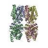
Journal: Nat Commun / Year: 2025 Title: Distinct oligomeric assemblies of STING induced by non-nucleotide agonists. Authors: Anant Gharpure / Ariana Sulpizio / Johannes R Loeffler / Monica L Fernández-Quintero / Andy S Tran / Luke L Lairson / Andrew B Ward / Abstract: STING plays essential roles coordinating innate immune responses to processes that range from pathogenic infection to genomic instability. Its adaptor function is activated by cyclic dinucleotide ...STING plays essential roles coordinating innate immune responses to processes that range from pathogenic infection to genomic instability. Its adaptor function is activated by cyclic dinucleotide (CDN) secondary messengers originating from self (2'3'-cGAMP) or bacterial sources (3'3'-CDNs). Different classes of CDNs possess distinct binding modes, stabilizing STING's ligand-binding domain (LBD) in either a closed or open conformation. The closed conformation, induced by the endogenous ligand 2'3'-cGAMP, has been extensively studied using cryo-EM. However, significant questions remain regarding the structural basis of STING activation by open conformation-inducing ligands. Using cryo-EM, we investigate potential differences in conformational changes and oligomeric assemblies of STING for closed and open conformation-inducing synthetic agonists. While we observe a characteristic 180° rotation for both classes, the open-LBD inducing agonist diABZI-3 uniquely induces a quaternary structure reminiscent but distinct from the reported autoinhibited state of apo-STING. Additionally, we observe slower rates of activation for this ligand class in functional assays, which collectively suggests the existence of a potential additional regulatory mechanism for open conformation-inducing ligands that involves head-to-head interactions and restriction of curved oligomer formation. These observations have potential implications in the selection of an optimal class of STING agonist in the context of a defined therapeutic application.
In the structure databanks used in Yorodumi, some data are registered as the other names, "COVID-19 virus" and "2019-nCoV". Here are the details of the virus and the list of structure data.
Jan 31, 2019. EMDB accession codes are about to change! (news from PDBe EMDB page)
EMDB accession codes are about to change! (news from PDBe EMDB page)
The allocation of 4 digits for EMDB accession codes will soon come to an end. Whilst these codes will remain in use, new EMDB accession codes will include an additional digit and will expand incrementally as the available range of codes is exhausted. The current 4-digit format prefixed with “EMD-” (i.e. EMD-XXXX) will advance to a 5-digit format (i.e. EMD-XXXXX), and so on. It is currently estimated that the 4-digit codes will be depleted around Spring 2019, at which point the 5-digit format will come into force.
The EM Navigator/Yorodumi systems omit the EMD- prefix.
Related info.:Q: What is EMD? / ID/Accession-code notation in Yorodumi/EM Navigator
Yorodumi is a browser for structure data from EMDB, PDB, SASBDB, etc.
This page is also the successor to EM Navigator detail page, and also detail information page/front-end page for Omokage search.
The word "yorodu" (or yorozu) is an old Japanese word meaning "ten thousand". "mi" (miru) is to see.
Related info.:EMDB / PDB / SASBDB / Comparison of 3 databanks / Yorodumi Search / Aug 31, 2016. New EM Navigator & Yorodumi / Yorodumi Papers / Jmol/JSmol / Function and homology information / Changes in new EM Navigator and Yorodumi
 Movie
Movie Controller


 Open data
 Basic information
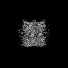
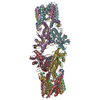
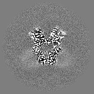
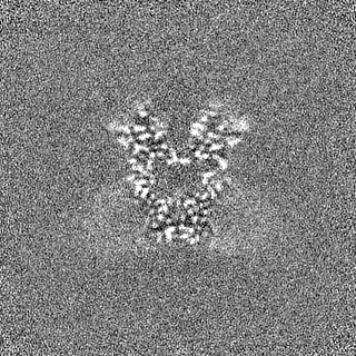
 Map data
 Sample
 Keywords
 Function and homology information
 Homo sapiens (human)
 Authors
 Citation
 Structure visualization
 Downloads & links
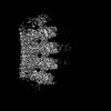
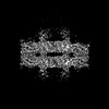
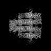
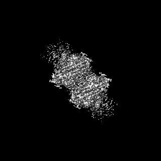
 emd_45897.png
 http://ftp.pdbj.org/pub/emdb/structures/EMD-45897
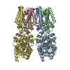


 Z (Sec.)
 Y (Row.)
 X (Col.)


 Sample components
 Processing
 Electron microscopy
 FIELD EMISSION GUN
