 Movie
Movie Controller

[English] 日本語
 Yorodumi
- EMDB-45591: Autoinhibited full-length LRRK2(I2020T) on microtubules with MLi-2 -
+ Open data

- Basic information
| Entry | 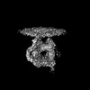 | |||||||||
|---|---|---|---|---|---|---|---|---|---|---|
| Title | Autoinhibited full-length LRRK2(I2020T) on microtubules with MLi-2 | |||||||||
 Map data Map data | IT_Mli2 | |||||||||
 Sample Sample |
| |||||||||
 Keywords Keywords | Kinase / GTPase / PROTEIN BINDING | |||||||||
| Function / homology |  Function and homology information Function and homology informationcaveola neck / : / beta-catenin destruction complex binding / regulation of branching morphogenesis of a nerve / Wnt signalosome assembly / negative regulation of motile cilium assembly / regulation of neuron maturation / regulation of kidney size / regulation of cell projection organization / regulation of dopamine receptor signaling pathway ...caveola neck / : / beta-catenin destruction complex binding / regulation of branching morphogenesis of a nerve / Wnt signalosome assembly / negative regulation of motile cilium assembly / regulation of neuron maturation / regulation of kidney size / regulation of cell projection organization / regulation of dopamine receptor signaling pathway / tangential migration from the subventricular zone to the olfactory bulb / GTP-dependent protein kinase activity / regulation of SNARE complex assembly / regulation of neuroblast proliferation / regulation of ER to Golgi vesicle-mediated transport / protein localization to endoplasmic reticulum exit site / negative regulation of late endosome to lysosome transport / regulation of mitochondrial depolarization / : / peroxidase inhibitor activity / positive regulation of dopamine receptor signaling pathway / amphisome / regulation of synaptic vesicle transport / : / regulation of CAMKK-AMPK signaling cascade / negative regulation of autophagosome assembly / co-receptor binding / positive regulation of microglial cell activation / regulation of retrograde transport, endosome to Golgi / negative regulation of GTPase activity / cellular response to curcumin / olfactory bulb development / regulation of locomotion / cytoplasmic side of mitochondrial outer membrane / positive regulation of synaptic vesicle endocytosis / JUN kinase kinase kinase activity / striatum development / multivesicular body, internal vesicle / negative regulation of excitatory postsynaptic potential / regulation of cAMP/PKA signal transduction / neuron projection arborization / regulation of dendritic spine morphogenesis / mitochondrion localization / endoplasmic reticulum organization / protein localization to mitochondrion / cellular response to dopamine / positive regulation of mitochondrial outer membrane permeabilization involved in apoptotic signaling pathway / negative regulation of protein processing / positive regulation of protein autoubiquitination / Wnt signalosome / positive regulation of programmed cell death / GTP metabolic process / regulation of canonical Wnt signaling pathway / regulation of reactive oxygen species metabolic process / syntaxin-1 binding / lysosome organization / negative regulation of macroautophagy / Golgi-associated vesicle / PTK6 promotes HIF1A stabilization / clathrin binding / Golgi organization / regulation of mitochondrial fission / Lewy body / regulation of synaptic vesicle exocytosis / intracellular distribution of mitochondria / exploration behavior / protein kinase A binding / microvillus / autolysosome / neuromuscular junction development / locomotory exploration behavior / endoplasmic reticulum exit site / canonical Wnt signaling pathway / MAP kinase kinase kinase activity / regulation of synaptic vesicle endocytosis / negative regulation of endoplasmic reticulum stress-induced intrinsic apoptotic signaling pathway / Rho protein signal transduction / regulation of synaptic transmission, glutamatergic / JNK cascade / presynaptic cytosol / cellular response to manganese ion / phagocytic vesicle / neuron projection morphogenesis / positive regulation of autophagy / dendrite cytoplasm / excitatory postsynaptic potential / determination of adult lifespan / positive regulation of protein ubiquitination / cellular response to starvation / regulation of autophagy / GTPase activator activity / SNARE binding / calcium-mediated signaling / mitochondrion organization / cellular response to reactive oxygen species / trans-Golgi network / regulation of protein stability / regulation of membrane potential / tubulin binding / autophagy Similarity search - Function | |||||||||
| Biological species |  Homo sapiens (human) Homo sapiens (human) | |||||||||
| Method | subtomogram averaging / cryo EM / Resolution: 7.8 Å | |||||||||
 Authors Authors | Chen S / Villa E / Leschziner AE | |||||||||
| Funding support |  United States, 2 items United States, 2 items
| |||||||||
 Citation Citation | Journal: bioRxiv / Year: 2025 Title: Cryo-electron tomography reveals the microtubule-bound form of inactive LRRK2. Authors: Siyu Chen / Tamar Basiashvili / Joshua Hutchings / Marta Sanz Murillo / Amalia Villagran Suarez / Erica Xiong / Jaime Alegrio Louro / Andres E Leschziner / Elizabeth Villa / Abstract: Parkinson's Disease (PD) is the second most common neurodegenerative disorder. Mutations in leucine-rich repeat kinase 2 (LRRK2), a multi-domain protein containing both a kinase and a GTPase, are a ...Parkinson's Disease (PD) is the second most common neurodegenerative disorder. Mutations in leucine-rich repeat kinase 2 (LRRK2), a multi-domain protein containing both a kinase and a GTPase, are a leading cause of the familial form of PD. Pathogenic LRRK2 mutations increase LRRK2 kinase activity. While the bulk of LRRK2 is found in the cytosol, the protein associates with membranes where its Rab GTPase substrates are found, and under certain conditions, with microtubules. Integrative structural studies using single-particle cryo-electron microscopy (cryo-EM) and cryo-electron tomography (cryo-ET) have revealed the architecture of microtubule-associated LRRK2 filaments, and that formation of these filaments requires LRRK2's kinase to be in the active-like conformation. However, whether LRRK2 can interact with and form filaments on microtubules in its autoinhibited state, where the kinase domain is in the inactive conformation and the N-terminal LRR domain covers the kinase active site, was not known. Using cryo-ET, we show that full-length LRRK2 can oligomerize on microtubules in its autoinhibited state. Both WT-LRRK2 and PD-linked LRRK2 mutants formed filaments on microtubules. While these filaments are stabilized by the same interfaces seen in the active-LRRK2 filaments, we observed a new interface involving the N-terminal repeats that were disordered in the active-LRRK2 filaments. The helical parameters of the autoinhibited-LRRK2 filaments are different from those reported for the active-LRRK2 filaments. Finally, the autoinhibited-LRRK2 filaments are shorter and less regular, suggesting they are less stable. #1: Journal: Elife / Year: 2024Title: Cryo-electron tomography reveals the microtubule-bound form of inactive LRRK2 Authors: Chen S / Basiashvili T / Hutchings J / Sanz Murillo M / Villagran Suarez A / Alegrio Louro J / Leschziner AE / Villa E | |||||||||
| History |
|
- Structure visualization
| Supplemental images |
|---|

- Downloads & links
-EMDB archive
| Map data | emd_45591.map.gz | 20 MB | EMDB map data format | |
|---|---|---|---|---|
| Header (meta data) | emd-45591-v30.xmlemd-45591.xml | 27.2 KB 27.2 KB | Display Display | EMDB header |
| FSC (resolution estimation) | emd_45591_fsc.xml | 6.9 KB | Display | FSC data file |
| Images |  emd_45591.png emd_45591.png | 82.4 KB | ||
| Filedesc metadata | emd-45591.cif.gz | 8.1 KB | ||
| Others | emd_45591_half_map_1.map.gzemd_45591_half_map_2.map.gz | 20.4 MB 20.4 MB | ||
| Archive directory |  http://ftp.pdbj.org/pub/emdb/structures/EMD-45591ftp://ftp.pdbj.org/pub/emdb/structures/EMD-45591 http://ftp.pdbj.org/pub/emdb/structures/EMD-45591ftp://ftp.pdbj.org/pub/emdb/structures/EMD-45591 | HTTPS FTP |
-Related structure data
| Related structure data | 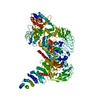 9choMC M: atomic model generated by this map C: citing same article ( |
|---|---|
| Similar structure data |


-Links
| EMDB pages | EMDB (EBI/PDBe) / EMDataResource |
|---|---|
| Related items in Molecule of the Month |


-Map
| File | Download / File: emd_45591.map.gz / Format: CCP4 / Size: 27 MB / Type: IMAGE STORED AS FLOATING POINT NUMBER (4 BYTES) | ||||||||||||||||||||||||||||||||||||
|---|---|---|---|---|---|---|---|---|---|---|---|---|---|---|---|---|---|---|---|---|---|---|---|---|---|---|---|---|---|---|---|---|---|---|---|---|---|
| Annotation | IT_Mli2 | ||||||||||||||||||||||||||||||||||||
| Projections & slices | Image control
Images are generated by Spider. | ||||||||||||||||||||||||||||||||||||
| Voxel size | X=Y=Z: 2.161 Å | ||||||||||||||||||||||||||||||||||||
| Density |
| ||||||||||||||||||||||||||||||||||||
| Symmetry | Space group: 1 | ||||||||||||||||||||||||||||||||||||
| Details | EMDB XML:
|
 Z (Sec.)
 Y (Row.)
 X (Col.)


-Supplemental data
-Half map: #2
| File | emd_45591_half_map_1.map | ||||||||||||
|---|---|---|---|---|---|---|---|---|---|---|---|---|---|
| Projections & Slices |
| ||||||||||||
| Density Histograms |


-Half map: #1
| File | emd_45591_half_map_2.map | ||||||||||||
|---|---|---|---|---|---|---|---|---|---|---|---|---|---|
| Projections & Slices |
| ||||||||||||
| Density Histograms |


- Sample components
-Entire : Full-length autoinhibited LRRK2 I2020T on microtubules
| Entire | Name: Full-length autoinhibited LRRK2 I2020T on microtubules |
|---|---|
| Components |
|
-Supramolecule #1: Full-length autoinhibited LRRK2 I2020T on microtubules
| Supramolecule | Name: Full-length autoinhibited LRRK2 I2020T on microtubules type: complex / ID: 1 / Parent: 0 / Macromolecule list: #1 |
|---|---|
| Source (natural) | Organism: Homo sapiens (human) |
| Molecular weight | Theoretical: 286.103 KDa |
-Macromolecule #1: Leucine-rich repeat serine/threonine-protein kinase 2
| Macromolecule | Name: Leucine-rich repeat serine/threonine-protein kinase 2 / type: protein_or_peptide / ID: 1 / Number of copies: 1 / Enantiomer: LEVO / EC number: non-specific serine/threonine protein kinase |
|---|---|
| Source (natural) | Organism: Homo sapiens (human) |
| Molecular weight | Theoretical: 224.904875 KDa |
| Recombinant expression | Organism:   Spodoptera frugiperda (fall armyworm) Spodoptera frugiperda (fall armyworm) |
| Sequence | String: HKLVLAALNR FIGNPGIQKC GLKVISSIVH FPDALEMLSL EGAMDSVLHT LQMYPDDQEI QCLGLSLIGY LITKKNVFIG TGHLLAKIL VSSLYRFKDV AEIQTKGFQT ILAILKLSAS FSKLLVHHSF DLVIFHQMSS NIMEQKDQQF LNLCCKCFAK V AMDDYLKN ...String: HKLVLAALNR FIGNPGIQKC GLKVISSIVH FPDALEMLSL EGAMDSVLHT LQMYPDDQEI QCLGLSLIGY LITKKNVFIG TGHLLAKIL VSSLYRFKDV AEIQTKGFQT ILAILKLSAS FSKLLVHHSF DLVIFHQMSS NIMEQKDQQF LNLCCKCFAK V AMDDYLKN VMLERACDQN NSIMVECLLL LGADANQAKE GSSLICQVCE KESSPKLVEL LLNSGSREQD VRKALTISIG KG DSQIISL LLRRLALDVA NNSICLGGFC IGKVEPSWLG PLFPDKTSNL RKQTNIASTL ARMVIRYQMK SAVEEGTASG SDG NFSEDV LSKFDEWTFI PDSSMDSVFA QSDDLDSEGS EGSFLVKKKS NSISVGEFYR DAVLQRCSPN LQRHSNSLGP IFDH EDLLK RKRKILSSDD SLRSSKLQSH MRHSDSISSL ASEREYITSL DLSANELRDI DALSQKCCIS VHLEHLEKLE LHQNA LTSF PQQLCETLKS LTHLDLHSNK FTSFPSYLLK MSCIANLDVS RNDIGPSVVL DPTVKCPTLK QFNLSYNQLS FVPENL TDV VEKLEQLILE GNKISGICSP LRLKELKILN LSKNHISSLS ENFLEACPKV ESFSARMNFL AAMPFLPPSM TILKLSQ NK FSCIPEAILN LPHLRSLDMS SNDIQYLPGP AHWKSLNLRE LLFSHNQISI LDLSEKAYLW SRVEKLHLSH NKLKEIPP E IGCLENLTSL DVSYNLELRS FPNEMGKLSK IWDLPLDELH LNFDFKHIGC KAKDIIRFLQ QRLKKAVPYN RMKLMIVGN TGSGKTTLLQ QLMKTKKSDL GMQSATVGID VKDWPIQIRD KRKRDLVLNV WDFAGREEFY STHPHFMTQR ALYLAVYDLS KGQAEVDAM KPWLFNIKAR ASSSPVILVG THLDVSDEKQ RKACMSKITK ELLNKRGFPA IRDYHFVNAT EESDALAKLR K TIINESLN FKIRDQLVVG QLIPDCYVEL EKIILSERKN VPIEFPVIDR KRLLQLVREN QLQLDENELP HAVHFLNESG VL LHFQDPA LQLSDLYFVE PKWLCKIMAQ ILTVKVEGCP KHPKGIISRR DVEKFLSKKR KFPKNYMSQY FKLLEKFQIA LPI GEEYLL VPSSLSDHRP VIELPHCENS EIIIRLYEMP YFPMGFWSRL INRLLEISPY MLSGRERALR PNRMYWRQGI YLNW SPEAY CLVGSEVLDN HPESFLKITV PSCRKGCILL GQVVDHIDSL MEEWFPGLLE IDICGEGETL LKKWALYSFN DGEEH QKIL LDDLMKKAEE GDLLVNPDQP RLTIPISQIA PDLILADLPR NIMLNNDELE FEQAPEFLLG DGSFGSVYRA AYEGEE VAV KIFNKHTSLR LLRQELVVLC HLHHPSLISL LAAGIRPRML VMELASKGSL DRLLQQDKAS LTRTLQHRIA LHVADGL RY LHSAMIIYRD LKPHNVLLFT LYPNAAIIAK IADYGTAQYC CRMGIKTSEG TPGFRAPEVA RGNVIYNQQA DVYSFGLL L YDILTTGGRI VEGLKFPNEF DELEIQGKLP DPVKEYGCAP WPMVEKLIKQ CLKENPQERP TSAQVFDILN SAELVCLTR RILLPKNVIV ECMVATHHNS RNASIWLGCG HTDRGQLSFL DLNTEGYTSE EVADSRILCL ALVHLPVEKE SWIVSGTQSG TLLVINTED GKKRHTLEKM TDSVTCLYCN SFSKQSKQKN FLLVGTADGK LAIFEDKTVK LKGAAPLKIL NIGNVSTPLM C LSESTNST ERNVMWGGCG TKIFSFSNDF TIQKLIETRT SQLFSYAAFS DSNIITVVVD TALYIAKQNS PVVEVWDKKT EK LCGLIDC VHFLREVMVK ENKESKHKMS YSGRVKTLCL QKNTALWIGT GGGHILLLDL STRRLIRVIY NFCNSVRVMM TAQ LGSLKN VMLVLGYNRK NTEGTQKQKE IQSCLTVWDI NLPHEVQNLE KHIEVRKELA EKMRRTSVE UniProtKB: Leucine-rich repeat serine/threonine-protein kinase 2 |
-Macromolecule #2: GUANOSINE-5'-DIPHOSPHATE
| Macromolecule | Name: GUANOSINE-5'-DIPHOSPHATE / type: ligand / ID: 2 / Number of copies: 1 / Formula: GDP |
|---|---|
| Molecular weight | Theoretical: 443.201 Da |
| Chemical component information |  ChemComp-GDP: |
-Macromolecule #3: (2~{R},6~{S})-2,6-dimethyl-4-[6-[5-(1-methylcyclopropyl)oxy-1~{H}...
| Macromolecule | Name: (2~{R},6~{S})-2,6-dimethyl-4-[6-[5-(1-methylcyclopropyl)oxy-1~{H}-indazol-3-yl]pyrimidin-4-yl]morpholine type: ligand / ID: 3 / Number of copies: 1 / Formula: A1N |
|---|---|
| Molecular weight | Theoretical: 379.456 Da |
| Chemical component information |  ChemComp-A1N: |
-Experimental details
-Structure determination
| Method | cryo EM |
|---|---|
 Processing Processing | subtomogram averaging |
| Aggregation state | filament |
-Sample preparation
| Concentration | 1.43 mg/mL | ||||||||||||||||||
|---|---|---|---|---|---|---|---|---|---|---|---|---|---|---|---|---|---|---|---|
| Buffer | pH: 7.4 Component:
| ||||||||||||||||||
| Grid | Model: EMS Lacey Carbon / Material: COPPER / Mesh: 300 / Support film - Material: CARBON / Support film - topology: CONTINUOUS / Pretreatment - Type: GLOW DISCHARGE / Pretreatment - Time: 60 sec. / Pretreatment - Atmosphere: AIR | ||||||||||||||||||
| Vitrification | Cryogen name: ETHANE / Chamber humidity: 100 % / Chamber temperature: 277.15 K / Instrument: FEI VITROBOT MARK IV |
- Electron microscopy
| Microscope | FEI TITAN KRIOS |
|---|---|
| Temperature | Min: 77.0 K / Max: 90.0 K |
| Software | Name: SerialEM (ver. 4.6) |
| Image recording | Film or detector model: GATAN K3 (6k x 4k) / Detector mode: COUNTING / Digitization - Dimensions - Width: 4092 pixel / Digitization - Dimensions - Height: 5760 pixel / Number real images: 1 / Average exposure time: 0.65 sec. / Average electron dose: 3.24 e/Å2 |
| Electron beam | Acceleration voltage: 300 kV / Electron source:  FIELD EMISSION GUN FIELD EMISSION GUN |
| Electron optics | C2 aperture diameter: 70.0 µm / Calibrated defocus max: 5.5 µm / Calibrated defocus min: 3.5 µm / Calibrated magnification: 42000 / Illumination mode: SPOT SCAN / Imaging mode: BRIGHT FIELD / Cs: 2.7 mm / Nominal defocus max: 5.0 µm / Nominal defocus min: 3.0 µm / Nominal magnification: 42000 |
| Sample stage | Specimen holder model: FEI TITAN KRIOS AUTOGRID HOLDER / Cooling holder cryogen: NITROGEN |
| Experimental equipment |  Model: Titan Krios / Image courtesy: FEI Company |
+Image processing

-Atomic model buiding 1
| Software | Name: UCSF ChimeraX (ver. 1.7) |
|---|---|
| Refinement | Space: REAL / Protocol: FLEXIBLE FIT |
| Output model | PDB-9cho: |
