 Movie
Movie Controller


+ Open data

- Basic information
| Entry |  | |||||||||
|---|---|---|---|---|---|---|---|---|---|---|
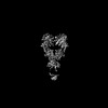
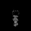
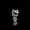
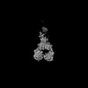
| Title | DosP Apo Bent form | |||||||||
 Map data Map data | ||||||||||
 Sample Sample |
| |||||||||
 Keywords Keywords | Heme / DosP / Phosphodiesterase / c-di-GMP / OXYGEN BINDING | |||||||||
| Function / homology |  Function and homology information Function and homology informationcyclic-guanylate-specific phosphodiesterase / regulation of single-species biofilm formation / cyclic-guanylate-specific phosphodiesterase activity / response to oxygen levels / oxygen sensor activity / heme binding / magnesium ion binding / protein homodimerization activity / plasma membrane Similarity search - Function | |||||||||
| Biological species |  | |||||||||
| Method | single particle reconstruction / cryo EM / Resolution: 3.97 Å | |||||||||
 Authors Authors | Kumar P / Kober DL | |||||||||
| Funding support |  United States, 2 items United States, 2 items
| |||||||||
 Citation Citation | Journal: Nat Commun / Year: 2024 Title: Structures of the multi-domain oxygen sensor DosP: remote control of a c-di-GMP phosphodiesterase by a regulatory PAS domain. Authors: Wenbi Wu / Pankaj Kumar / Chad A Brautigam / Shih-Chia Tso / Hamid R Baniasadi / Daniel L Kober / Marie-Alda Gilles-Gonzalez / Abstract: The heme-based direct oxygen sensor DosP degrades c-di-GMP, a second messenger nearly unique to bacteria. In stationary phase Escherichia coli, DosP is the most abundant c-di-GMP phosphodiesterase. ...The heme-based direct oxygen sensor DosP degrades c-di-GMP, a second messenger nearly unique to bacteria. In stationary phase Escherichia coli, DosP is the most abundant c-di-GMP phosphodiesterase. Ligation of O to a heme-binding PAS domain (hPAS) of the protein enhances the phosphodiesterase through an allosteric mechanism that has remained elusive. We determine six structures of full-length DosP in its aerobic or anaerobic conformations, with or without c-di-GMP. DosP is an elongated dimer with the regulatory heme containing domain and phosphodiesterase separated by nearly 180 Å. In the absence of substrate, regardless of the heme status, DosP presents an equilibrium of two distinct conformations. Binding of substrate induces DosP to adopt a single, ON-state or OFF-state conformation depending on its heme status. Structural and biochemical studies of this multi-domain sensor and its mutants provide insights into signal regulation of second-messenger levels. | |||||||||
| History |
|
- Structure visualization
| Supplemental images |
|---|

- Downloads & links
-EMDB archive
| Map data | emd_45489.map.gz | 32.4 MB | EMDB map data format | |
|---|---|---|---|---|
| Header (meta data) | emd-45489-v30.xmlemd-45489.xml | 20.4 KB 20.4 KB | Display Display | EMDB header |
| FSC (resolution estimation) | emd_45489_fsc.xml | 8.4 KB | Display | FSC data file |
| Images |  emd_45489.png emd_45489.png | 28.9 KB | ||
| Filedesc metadata | emd-45489.cif.gz | 6.6 KB | ||
| Others | emd_45489_additional_1.map.gzemd_45489_half_map_1.map.gzemd_45489_half_map_2.map.gz | 57.2 MB 59.3 MB 59.3 MB | ||
| Archive directory |  http://ftp.pdbj.org/pub/emdb/structures/EMD-45489ftp://ftp.pdbj.org/pub/emdb/structures/EMD-45489 http://ftp.pdbj.org/pub/emdb/structures/EMD-45489ftp://ftp.pdbj.org/pub/emdb/structures/EMD-45489 | HTTPS FTP |
-Validation report
| Summary document | emd_45489_validation.pdf.gz | 721.4 KB | Display | EMDB validaton report |
|---|---|---|---|---|
| Full document | emd_45489_full_validation.pdf.gz | 721 KB | Display | |
| Data in XML | emd_45489_validation.xml.gz | 16.3 KB | Display | |
| Data in CIF | emd_45489_validation.cif.gz | 21.4 KB | Display | |
| Arichive directory | https://ftp.pdbj.org/pub/emdb/validation_reports/EMD-45489ftp://ftp.pdbj.org/pub/emdb/validation_reports/EMD-45489 | HTTPS FTP |
-Related structure data
| Related structure data |  9ce0MC  9bgvC  9bkvC  9cdrC  9cloC  9cmfC M: atomic model generated by this map C: citing same article ( |
|---|---|
| Similar structure data |


-Links
| EMDB pages | EMDB (EBI/PDBe) / EMDataResource |
|---|---|
| Related items in Molecule of the Month |


-Map
| File | Download / File: emd_45489.map.gz / Format: CCP4 / Size: 64 MB / Type: IMAGE STORED AS FLOATING POINT NUMBER (4 BYTES) | ||||||||||||||||||||||||||||||||||||
|---|---|---|---|---|---|---|---|---|---|---|---|---|---|---|---|---|---|---|---|---|---|---|---|---|---|---|---|---|---|---|---|---|---|---|---|---|---|
| Projections & slices | Image control
Images are generated by Spider. | ||||||||||||||||||||||||||||||||||||
| Voxel size | X=Y=Z: 1.61445 Å | ||||||||||||||||||||||||||||||||||||
| Density |
| ||||||||||||||||||||||||||||||||||||
| Symmetry | Space group: 1 | ||||||||||||||||||||||||||||||||||||
| Details | EMDB XML:
|
 Z (Sec.)
 Y (Row.)
 X (Col.)


-Supplemental data
-Additional map: #1
| File | emd_45489_additional_1.map | ||||||||||||
|---|---|---|---|---|---|---|---|---|---|---|---|---|---|
| Projections & Slices |
| ||||||||||||
| Density Histograms |


-Half map: #2
| File | emd_45489_half_map_1.map | ||||||||||||
|---|---|---|---|---|---|---|---|---|---|---|---|---|---|
| Projections & Slices |
| ||||||||||||
| Density Histograms |


-Half map: #1
| File | emd_45489_half_map_2.map | ||||||||||||
|---|---|---|---|---|---|---|---|---|---|---|---|---|---|
| Projections & Slices |
| ||||||||||||
| Density Histograms |


- Sample components
-Entire : DosP apo bent form
| Entire | Name: DosP apo bent form |
|---|---|
| Components |
|
-Supramolecule #1: DosP apo bent form
| Supramolecule | Name: DosP apo bent form / type: complex / ID: 1 / Parent: 0 / Macromolecule list: #1 |
|---|---|
| Source (natural) | Organism: |
| Molecular weight | Theoretical: 200 KDa |
-Macromolecule #1: Oxygen sensor protein DosP
| Macromolecule | Name: Oxygen sensor protein DosP / type: protein_or_peptide / ID: 1 / Number of copies: 2 / Enantiomer: LEVO / EC number: cyclic-guanylate-specific phosphodiesterase |
|---|---|
| Source (natural) | Organism: |
| Molecular weight | Theoretical: 91.154109 KDa |
| Recombinant expression | Organism: |
| Sequence | String: MRQDAEVIMK LTDADSAADG IFFPALEQNM MGAVLINEND EVMFFNPAAE KLWGYKREEV IGNNIDMLIP RDLRPAHPEY IRHNREGGK ARVEGMSREL QLEKKDGSKI WTRFALSKVS AEGKVYYLAL VRDASVEMAQ KEQTRQLIIA VDHLDRPVIV L DPERHIVQ ...String: MRQDAEVIMK LTDADSAADG IFFPALEQNM MGAVLINEND EVMFFNPAAE KLWGYKREEV IGNNIDMLIP RDLRPAHPEY IRHNREGGK ARVEGMSREL QLEKKDGSKI WTRFALSKVS AEGKVYYLAL VRDASVEMAQ KEQTRQLIIA VDHLDRPVIV L DPERHIVQ CNRAFTEMFG YCISEASGMQ PDTLLNTPEF PADNRIRLQQ LLWKTARDQD EFLLLTRTGE KIWIKASISP VY DVLAHLQ NLVMTFSDIT EERQIRQLEG NILAAMCSSP PFHEMGEIIC RNIESVLNES HVSLFALRNG MPIHWASSSH GAE IQNAQS WSATIRQRDG APAGILQIKT SSGAETSAFI ERVADISQHM AALALEQEKS RQHIEQLIQF DPMTGLPNRN NLHN YLDDL VDKAVSPVVY LIGVDHIQDV IDSLGYAWAD QALLEVVNRF REKLKPDQYL CRIEGTQFVL VSLENDVSNI TQIAD ELRN VVSKPIMIDD KPFPLTLSIG ISYDLGKNRD YLLSTAHNAM DYIRKNGGNG WQFFSPAMNE MVKERLVLGA ALKEAI SNN QLKLVYQPQI FAETGELYGI EALARWHDPL HGHVPPSRFI PLAEEIGEIE NIGRWVIAEA CRQLAEWRSQ NIHIPAL SV NLSALHFRSN QLPNQVSDAM HAWGIDGHQL TVEITESMMM EHDTEIFKRI QILRDMGVGL SVDDFGTGFS GLSRLVSL P VTEIKIDKSF VDRCLTEKRI LALLEAITSI GQSLNLTVVA EGVETKEQFE MLRKIHCRVI QGYFFSRPLP AEEIPGWMS SVLPLK UniProtKB: Oxygen sensor protein DosP |
-Macromolecule #2: PROTOPORPHYRIN IX CONTAINING FE
| Macromolecule | Name: PROTOPORPHYRIN IX CONTAINING FE / type: ligand / ID: 2 / Number of copies: 2 / Formula: HEM |
|---|---|
| Molecular weight | Theoretical: 616.487 Da |
| Chemical component information |  ChemComp-HEM: |
-Macromolecule #3: OXYGEN MOLECULE
| Macromolecule | Name: OXYGEN MOLECULE / type: ligand / ID: 3 / Number of copies: 2 / Formula: OXY |
|---|---|
| Molecular weight | Theoretical: 31.999 Da |
| Chemical component information |  ChemComp-O2: |
-Experimental details
-Structure determination
| Method | cryo EM |
|---|---|
 Processing Processing | single particle reconstruction |
| Aggregation state | particle |
-Sample preparation
| Buffer | pH: 7.5 |
|---|---|
| Grid | Model: Quantifoil R1.2/1.3 / Material: GOLD / Mesh: 300 / Pretreatment - Type: GLOW DISCHARGE / Pretreatment - Time: 80 sec. / Pretreatment - Atmosphere: AIR |
| Vitrification | Cryogen name: ETHANE |
- Electron microscopy
| Microscope | TFS KRIOS |
|---|---|
| Image recording | Film or detector model: GATAN K3 BIOQUANTUM (6k x 4k) / Average electron dose: 50.0 e/Å2 |
| Electron beam | Acceleration voltage: 300 kV / Electron source:  FIELD EMISSION GUN FIELD EMISSION GUN |
| Electron optics | Illumination mode: FLOOD BEAM / Imaging mode: BRIGHT FIELD / Nominal defocus max: 2.0 µm / Nominal defocus min: 0.8 µm |
| Experimental equipment |  Model: Titan Krios / Image courtesy: FEI Company |

