National Institutes of Health/National Institute of Arthritis and Musculoskeletal and Skin Diseases (NIH/NIAMS)
Intramural Research Program
United States
Citation
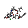
Journal: J Biol Chem / Year: 2025 Title: Structure of blood cell-specific tubulin and demonstration of dimer spacing compaction in a single protofilament. Authors: Felipe Montecinos / Elif Eren / Norman R Watts / Dan L Sackett / Paul T Wingfield / Abstract: Microtubule (MT) function plasticity originates from its composition of α- and β-tubulin isotypes and the posttranslational modifications of both subunits. Aspects such as MT assembly dynamics, ...Microtubule (MT) function plasticity originates from its composition of α- and β-tubulin isotypes and the posttranslational modifications of both subunits. Aspects such as MT assembly dynamics, structure, and anticancer drug binding can be modulated by αβ-tubulin heterogeneity. However, the exact molecular mechanism regulating these aspects is only partially understood. A recent insight is the discovery of expansion and compaction of the MT lattice, which can occur via fine modulation of dimer longitudinal spacing mediated by GTP hydrolysis, taxol binding, protein binding, or isotype composition. Here, we report the first structure of the blood cell-specific α1/β1-tubulin isolated from the marginal band of chicken erythrocytes (ChET) determined to a resolution of 3.2 Å by cryo-EM. We show that ChET rings induced with cryptophycin-52 (Cp-52) are smaller in diameter than HeLa cell line tubulin (HeLaT) rings induced with Cp-52 and composed of the same number of heterodimers. We observe compacted interdimer and intradimer curved protofilament interfaces, characterized by shorter distances between ChET subunits and accompanied by conformational changes in the β-tubulin subunit. The compacted ChET interdimer interface brings more residues near the Cp-52 binding site. We measured the Cp-52 apparent binding affinities of ChET and HeLaT by mass photometry, observing small differences, and detected the intermediates of the ring assembly reaction. These findings demonstrate that compaction/expansion of dimer spacing can occur in a single protofilament context and that the subtle structural differences between tubulin isotypes can modulate tubulin small molecule binding.
Model: Quantifoil R1.2/1.3 / Material: COPPER / Mesh: 400 / Support film - Material: CARBON / Support film - topology: CONTINUOUS / Support film - Film thickness: 2 / Pretreatment - Type: GLOW DISCHARGE / Pretreatment - Time: 15 sec. / Pretreatment - Atmosphere: AIR
In the structure databanks used in Yorodumi, some data are registered as the other names, "COVID-19 virus" and "2019-nCoV". Here are the details of the virus and the list of structure data.
Jan 31, 2019. EMDB accession codes are about to change! (news from PDBe EMDB page)
EMDB accession codes are about to change! (news from PDBe EMDB page)
The allocation of 4 digits for EMDB accession codes will soon come to an end. Whilst these codes will remain in use, new EMDB accession codes will include an additional digit and will expand incrementally as the available range of codes is exhausted. The current 4-digit format prefixed with “EMD-” (i.e. EMD-XXXX) will advance to a 5-digit format (i.e. EMD-XXXXX), and so on. It is currently estimated that the 4-digit codes will be depleted around Spring 2019, at which point the 5-digit format will come into force.
The EM Navigator/Yorodumi systems omit the EMD- prefix.
Related info.:Q: What is EMD? / ID/Accession-code notation in Yorodumi/EM Navigator
Yorodumi is a browser for structure data from EMDB, PDB, SASBDB, etc.
This page is also the successor to EM Navigator detail page, and also detail information page/front-end page for Omokage search.
The word "yorodu" (or yorozu) is an old Japanese word meaning "ten thousand". "mi" (miru) is to see.
Related info.:EMDB / PDB / SASBDB / Comparison of 3 databanks / Yorodumi Search / Aug 31, 2016. New EM Navigator & Yorodumi / Yorodumi Papers / Jmol/JSmol / Function and homology information / Changes in new EM Navigator and Yorodumi
 Movie
Movie Controller

 Yorodumi
 Open data
 Basic information
 Map data
 Sample
 Keywords
 Function and homology information
 Authors
 United States, 1 items
  Citation
 Structure visualization
 Downloads & links
 emd_45265.png
 http://ftp.pdbj.org/pub/emdb/structures/EMD-45265


 Z (Sec.)
 Y (Row.)
 X (Col.)


 Sample components

 Processing
 Electron microscopy
 FIELD EMISSION GUN

