National Institutes of Health/National Heart, Lung, and Blood Institute (NIH/NHLBI)
R00HL138129
United States
National Institutes of Health/National Institute of General Medical Sciences (NIH/NIGMS)
DP2GM149551
United States
Citation
Journal: Sci Adv / Year: 2025 Title: Human P2X4 receptor gating is modulated by a stable cytoplasmic cap and a unique allosteric pocket. Authors: Haoyuan Shi / Ismayn A Ditter / Adam C Oken / Steven E Mansoor / Abstract: P2X receptors (P2XRs) are adenosine 5'-triphosphate (ATP)-gated ion channels comprising homomeric and heteromeric trimers of seven subtypes (P2X1-P2X7) that confer different rates of desensitization. ...P2X receptors (P2XRs) are adenosine 5'-triphosphate (ATP)-gated ion channels comprising homomeric and heteromeric trimers of seven subtypes (P2X1-P2X7) that confer different rates of desensitization. The helical recoil model of P2XR desensitization proposes stability of the cytoplasmic cap sets the rate of desensitization, but timing of its formation is unclear for slow-desensitizing P2XRs. We report cryo-electron microscopy structures of full-length wild-type human P2X4 receptor in apo closed, antagonist-bound inhibited, and ATP-bound desensitized states. Because the apo closed and antagonist-bound inhibited state structures of this slow-desensitizing P2XR include an intact cytoplasmic cap while the ATP-bound desensitized state structure does not, the cytoplasmic cap is formed before agonist binding. Furthermore, structural and functional data suggest the cytoplasmic cap is stabilized by lipids to modulate desensitization, and P2X4 is modified by glycosylation and palmitoylation. Last, our antagonist-bound inhibited state structure reveals features specific to the allosteric ligand-binding pocket in human receptors that facilitates development of small-molecule modulators.
In the structure databanks used in Yorodumi, some data are registered as the other names, "COVID-19 virus" and "2019-nCoV". Here are the details of the virus and the list of structure data.
Jan 31, 2019. EMDB accession codes are about to change! (news from PDBe EMDB page)
EMDB accession codes are about to change! (news from PDBe EMDB page)
The allocation of 4 digits for EMDB accession codes will soon come to an end. Whilst these codes will remain in use, new EMDB accession codes will include an additional digit and will expand incrementally as the available range of codes is exhausted. The current 4-digit format prefixed with “EMD-” (i.e. EMD-XXXX) will advance to a 5-digit format (i.e. EMD-XXXXX), and so on. It is currently estimated that the 4-digit codes will be depleted around Spring 2019, at which point the 5-digit format will come into force.
The EM Navigator/Yorodumi systems omit the EMD- prefix.
Related info.:Q: What is EMD? / ID/Accession-code notation in Yorodumi/EM Navigator
Yorodumi is a browser for structure data from EMDB, PDB, SASBDB, etc.
This page is also the successor to EM Navigator detail page, and also detail information page/front-end page for Omokage search.
The word "yorodu" (or yorozu) is an old Japanese word meaning "ten thousand". "mi" (miru) is to see.
Related info.:EMDB / PDB / SASBDB / Comparison of 3 databanks / Yorodumi Search / Aug 31, 2016. New EM Navigator & Yorodumi / Yorodumi Papers / Jmol/JSmol / Function and homology information / Changes in new EM Navigator and Yorodumi
 Movie
Movie Controller

 Yorodumi
 Open data
 Basic information
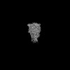
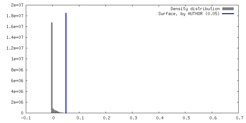
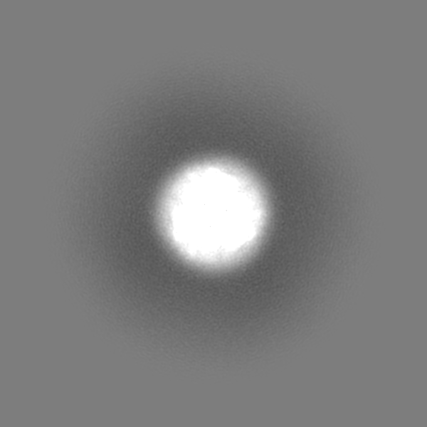
 Map data
 Sample
 Keywords
 Function and homology information
 Homo sapiens (human)
 Authors
 United States, 2 items
  Citation
 Structure visualization
 Downloads & links
 emd_45177.png
 http://ftp.pdbj.org/pub/emdb/structures/EMD-45177


 Z (Sec.)
 Y (Row.)
 X (Col.)


 Sample components


 Processing
 Electron microscopy
 FIELD EMISSION GUN


