Movie
Movie Controller


+ Open data

- Basic information
| Entry |  | |||||||||
|---|---|---|---|---|---|---|---|---|---|---|
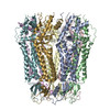
| Title | Pannexin 1 lacking C-terminal activating domain | |||||||||
 Map data Map data | ||||||||||
 Sample Sample |
| |||||||||
 Keywords Keywords | ATP release channel / nanodisc / large-pore / C-terminal activating domain / MEMBRANE PROTEIN | |||||||||
| Function / homology |  Function and homology information Function and homology informationpositive regulation of interleukin-1 production / gap junction / monoatomic cation transport / channel activity / monoatomic ion transmembrane transport / endoplasmic reticulum membrane / plasma membrane Similarity search - Function | |||||||||
| Biological species | ||||||||||
| Method | single particle reconstruction / cryo EM / Resolution: 2.9 Å | |||||||||
 Authors Authors | Ehrlich JJ / Kawate T | |||||||||
| Funding support |  United States, 1 items United States, 1 items
| |||||||||
 Citation Citation | Journal: Proc Natl Acad Sci U S A / Year: 2024 Title: The C-terminal activating domain promotes pannexin 1 channel opening. Authors: Erik Henze / Jacqueline J Ehrlich / Janice L Robertson / Eric Gelsleichter / Toshimitsu Kawate / Abstract: Pannexin 1 (Panx1) constitutes a large pore channel responsible for the release of adenosine triphosphate (ATP) from apoptotic cells. Strong evidence indicates that caspase-mediated cleavage of the C- ...Pannexin 1 (Panx1) constitutes a large pore channel responsible for the release of adenosine triphosphate (ATP) from apoptotic cells. Strong evidence indicates that caspase-mediated cleavage of the C-terminus promotes the opening of the Panx1 channel by unplugging the pore. However, this simple pore-plugging mechanism alone cannot account for the observation that a Panx1 construct ending before the caspase cleavage site remains closed. Here, we show that a helical region located immediately before the caspase cleavage site, referred to as the "C-terminal activating domain (CAD)", plays a pivotal role in facilitating Panx1 activation. Electrophysiology and mutagenesis studies uncovered that two conserved leucine residues within the CAD play a pivotal role. Cryoelectron microscopy (Cryo-EM) analysis of the construct ending before reaching the CAD demonstrated that the N terminus extends into an intracellular pocket. In contrast, the construct including the CAD revealed that this domain occupies the intracellular pocket, causing the N terminus to flip upward within the pore. Analysis of electrostatic free energy landscape in the closed conformation indicated that the intracellular side of the ion permeation pore may be occupied by anions like ATP, creating an electrostatic barrier for anions attempting to permeate the pore. When the N terminus flips up, it diminishes the positively charged surface, thereby reducing the drive to accumulate anions inside the pore. This dynamic change in the electrostatic landscape likely contributes to the selection of permeant ions. Collectively, these experiments put forth a mechanism in which C-terminal cleavage liberates the CAD, causing the repositioning of the N terminus to promote Panx1 channel opening. | |||||||||
| History |
|
- Structure visualization
| Supplemental images |
|---|

- Downloads & links
-EMDB archive
| Map data | emd_45055.map.gz | 31.1 MB | EMDB map data format | |
|---|---|---|---|---|
| Header (meta data) | emd-45055-v30.xmlemd-45055.xml | 18.2 KB 18.2 KB | Display Display | EMDB header |
| FSC (resolution estimation) | emd_45055_fsc.xml | 7.4 KB | Display | FSC data file |
| Images |  emd_45055.png emd_45055.png | 135.3 KB | ||
| Masks | emd_45055_msk_1.map | 274.6 MB | Mask map | |
| Filedesc metadata | emd-45055.cif.gz | 6.7 KB | ||
| Others | emd_45055_half_map_1.map.gzemd_45055_half_map_2.map.gz | 26.4 MB 26.4 MB | ||
| Archive directory |  http://ftp.pdbj.org/pub/emdb/structures/EMD-45055ftp://ftp.pdbj.org/pub/emdb/structures/EMD-45055 http://ftp.pdbj.org/pub/emdb/structures/EMD-45055ftp://ftp.pdbj.org/pub/emdb/structures/EMD-45055 | HTTPS FTP |
-Related structure data
| Related structure data |  9bz7MC  9bz8C M: atomic model generated by this map C: citing same article ( |
|---|---|
| Similar structure data |

-Links
| EMDB pages | EMDB (EBI/PDBe) / EMDataResource |
|---|
-Map
| File | Download / File: emd_45055.map.gz / Format: CCP4 / Size: 34.3 MB / Type: IMAGE STORED AS FLOATING POINT NUMBER (4 BYTES) | ||||||||||||||||||||||||||||||||||||
|---|---|---|---|---|---|---|---|---|---|---|---|---|---|---|---|---|---|---|---|---|---|---|---|---|---|---|---|---|---|---|---|---|---|---|---|---|---|
| Projections & slices | Image control
Images are generated by Spider. | ||||||||||||||||||||||||||||||||||||
| Voxel size | X=Y=Z: 1.07 Å | ||||||||||||||||||||||||||||||||||||
| Density |
| ||||||||||||||||||||||||||||||||||||
| Symmetry | Space group: 1 | ||||||||||||||||||||||||||||||||||||
| Details | EMDB XML:
|
 Z (Sec.)
 Y (Row.)
 X (Col.)


-Supplemental data
-Mask #1
| File | emd_45055_msk_1.map | ||||||||||||
|---|---|---|---|---|---|---|---|---|---|---|---|---|---|
| Projections & Slices |
| ||||||||||||
| Density Histograms |


-Half map: #2
| File | emd_45055_half_map_1.map | ||||||||||||
|---|---|---|---|---|---|---|---|---|---|---|---|---|---|
| Projections & Slices |
| ||||||||||||
| Density Histograms |


-Half map: #1
| File | emd_45055_half_map_2.map | ||||||||||||
|---|---|---|---|---|---|---|---|---|---|---|---|---|---|
| Projections & Slices |
| ||||||||||||
| Density Histograms |


- Sample components
-Entire : frog pannexin 1
| Entire | Name: frog pannexin 1 |
|---|---|
| Components |
|
-Supramolecule #1: frog pannexin 1
| Supramolecule | Name: frog pannexin 1 / type: complex / ID: 1 / Parent: 0 / Macromolecule list: all Details: Engineered frog pannexin 1 truncated after amino acid 357 in lipid nanodiscs |
|---|---|
| Source (natural) | Organism: |
-Macromolecule #1: Pannexin
| Macromolecule | Name: Pannexin / type: protein_or_peptide / ID: 1 / Number of copies: 7 / Enantiomer: LEVO |
|---|---|
| Source (natural) | Organism: |
| Molecular weight | Theoretical: 39.757516 KDa |
| Recombinant expression | Organism:  Trichoplusia ni (cabbage looper) Trichoplusia ni (cabbage looper) |
| Sequence | String: VFSDFLLKDP PESKYKGLRL ELAVDKLVSC IAVGLPLLLI SLAFAQEITL GSQISCFAPT SFSWRQAAYV DSFCWAAVQQ KHLSQSDSG NVPLWLHKFF PYILLLVAVL LYLPNLFWRF TAAPHLSSDL KFVMEELDKC YNRDIKDIKA ANNLNSSDKR D GLNSPVVS ...String: VFSDFLLKDP PESKYKGLRL ELAVDKLVSC IAVGLPLLLI SLAFAQEITL GSQISCFAPT SFSWRQAAYV DSFCWAAVQQ KHLSQSDSG NVPLWLHKFF PYILLLVAVL LYLPNLFWRF TAAPHLSSDL KFVMEELDKC YNRDIKDIKA ANNLNSSDKR D GLNSPVVS ENLQQSLWEI PLSHYKYPIV EQYLKTKNNS YGLIIKYLIC RVVTLIIVFT ACIYLGYYIS LFSLTDEFTC NI RTGILRN DTALPPLVQC KLIAVGVFRL LSYINLIIYV LIMPFIIYAM LVPFRKTANV LKVYEVLPTF SVQQAPSKTY DDH SLFLLF LEENVSELKS YKFLKVLENI K UniProtKB: Pannexin |
-Experimental details
-Structure determination
| Method | cryo EM |
|---|---|
 Processing Processing | single particle reconstruction |
| Aggregation state | particle |
-Sample preparation
| Concentration | 1.5 mg/mL | |||||||||
|---|---|---|---|---|---|---|---|---|---|---|
| Buffer | pH: 7.4 Component:
Details: Sample was subject to size exclusion chromatography with HEPES/NaCl buffer. | |||||||||
| Grid | Model: Quantifoil R1.2/1.3 / Material: GOLD / Mesh: 300 / Support film - Material: CARBON / Support film - topology: HOLEY / Support film - Film thickness: 45 / Pretreatment - Type: GLOW DISCHARGE / Pretreatment - Time: 180 sec. / Pretreatment - Atmosphere: AIR | |||||||||
| Vitrification | Cryogen name: ETHANE / Chamber humidity: 100 % / Chamber temperature: 288.15 K / Instrument: FEI VITROBOT MARK IV Details: Grid was blotted for 4 seconds with a force of 7 before plunging into liquid ethane.. | |||||||||
| Details | frPanx1 in lipid nanodiscs containing POPC, POPG, POPE and cholesterol. Sample eluted in a single peak from size-exclusion and was pooled and concentrated. |
- Electron microscopy
| Microscope | FEI TALOS ARCTICA |
|---|---|
| Specialist optics | Energy filter - Name: GIF Bioquantum / Energy filter - Slit width: 15 eV |
| Image recording | Film or detector model: GATAN K3 BIOQUANTUM (6k x 4k) / Number grids imaged: 1 / Number real images: 8713 / Average exposure time: 3.81 sec. / Average electron dose: 50.0 e/Å2 Details: 50-frame movies were collected in counted super resolution mode. |
| Electron beam | Acceleration voltage: 300 kV / Electron source:  FIELD EMISSION GUN FIELD EMISSION GUN |
| Electron optics | C2 aperture diameter: 100.0 µm / Illumination mode: FLOOD BEAM / Imaging mode: BRIGHT FIELD / Cs: 2.7 mm / Nominal defocus max: 20.0 µm / Nominal defocus min: 8.0 µm / Nominal magnification: 81000 |
| Sample stage | Specimen holder model: FEI TITAN KRIOS AUTOGRID HOLDER / Cooling holder cryogen: NITROGEN |
| Experimental equipment |  Model: Talos Arctica / Image courtesy: FEI Company |