 Movie
Movie Controller


+ Open data

- Basic information
| Entry |  | |||||||||
|---|---|---|---|---|---|---|---|---|---|---|
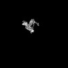
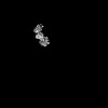
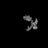
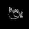
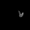
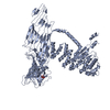
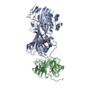
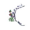
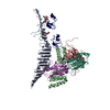
| Title | Dimeric form of LDL, LDL receptor and Legobody | |||||||||
 Map data Map data | ||||||||||
 Sample Sample |
| |||||||||
 Keywords Keywords | Apolipoprotein B 100 / ApoB 100 / LDL / LDL receptor / LIPID TRANSPORT | |||||||||
| Biological species |  Homo sapiens (human) / Homo sapiens (human) /   | |||||||||
| Method | single particle reconstruction / cryo EM / Resolution: 8.6 Å | |||||||||
 Authors Authors | Dearborn AD / Reimund M / Graziano G / Lei H / Kumar A / Neufeld EB / Remaley AT / Marcotrigiano J | |||||||||
| Funding support |  United States, 1 items United States, 1 items
| |||||||||
 Citation Citation | Journal: Nature / Year: 2025 Title: Structure of apolipoprotein B100 bound to the low-density lipoprotein receptor. Authors: Mart Reimund / Altaira D Dearborn / Giorgio Graziano / Haotian Lei / Anthony M Ciancone / Ashish Kumar / Ronald Holewinski / Edward B Neufeld / Francis J O'Reilly / Alan T Remaley / Joseph Marcotrigiano / Abstract: Apolipoprotein B100 (apoB100) is a structural component of low-density lipoprotein (LDL) and a ligand for the LDL receptor (LDLR). Mutations in apoB100 or in LDLR cause familial ...Apolipoprotein B100 (apoB100) is a structural component of low-density lipoprotein (LDL) and a ligand for the LDL receptor (LDLR). Mutations in apoB100 or in LDLR cause familial hypercholesterolaemia, an autosomal dominant disease that is characterized by a marked increase in LDL cholesterol (LDL-C) and a higher risk of cardiovascular disease. The structure of apoB100 on LDL and its interaction with LDLR are poorly understood. Here we present the cryo-electron microscopy structures of apoB100 on LDL bound to the LDLR and a nanobody complex, which can form a C-symmetric, higher-order complex. Using local refinement, we determined high-resolution structures of the interfaces between apoB100 and LDLR. One binding interface is formed between several small-ligand-binding modules of LDLR and a series of basic patches that are scattered along a β-belt formed by apoB100, encircling LDL. The other binding interface is formed between the β-propeller domain of LDLR and the N-terminal domain of apoB100. Our results reveal how both interfaces are involved in LDL dimer formation, and how LDLR cycles between LDL- and self-bound conformations. In addition, known mutations in either apoB100 or LDLR, associated with high levels of LDL-C, are located at the LDL-LDLR interface. | |||||||||
| History |
|
- Structure visualization
| Supplemental images |
|---|

- Downloads & links
-EMDB archive
| Map data | emd_44442.map.gz | 482.9 MB |  EMDB map data format EMDB map data format | |
|---|---|---|---|---|
| Header (meta data) | emd-44442-v30.xmlemd-44442.xml | 16.1 KB 16.1 KB | Display Display | EMDB header |
| FSC (resolution estimation) | emd_44442_fsc.xml | 16.9 KB | Display | FSC data file |
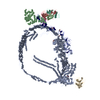
| Images |  emd_44442.png emd_44442.png | 74.6 KB | ||
| Filedesc metadata | emd-44442.cif.gz | 4.6 KB | ||
| Others | emd_44442_half_map_1.map.gzemd_44442_half_map_2.map.gz | 475.1 MB 475.1 MB | ||
| Archive directory |  http://ftp.pdbj.org/pub/emdb/structures/EMD-44442ftp://ftp.pdbj.org/pub/emdb/structures/EMD-44442 http://ftp.pdbj.org/pub/emdb/structures/EMD-44442ftp://ftp.pdbj.org/pub/emdb/structures/EMD-44442 | HTTPS FTP |
-Related structure data
-Links
| EMDB pages | EMDB (EBI/PDBe) / EMDataResource |
|---|
-Map
| File | Download / File: emd_44442.map.gz / Format: CCP4 / Size: 512 MB / Type: IMAGE STORED AS FLOATING POINT NUMBER (4 BYTES) | ||||||||||||||||||||||||||||||||||||
|---|---|---|---|---|---|---|---|---|---|---|---|---|---|---|---|---|---|---|---|---|---|---|---|---|---|---|---|---|---|---|---|---|---|---|---|---|---|
| Projections & slices | Image control
Images are generated by Spider. | ||||||||||||||||||||||||||||||||||||
| Voxel size | X=Y=Z: 1.66 Å | ||||||||||||||||||||||||||||||||||||
| Density |
| ||||||||||||||||||||||||||||||||||||
| Symmetry | Space group: 1 | ||||||||||||||||||||||||||||||||||||
| Details | EMDB XML:
|
 Z (Sec.)
 Y (Row.)
 X (Col.)


-Supplemental data
-Half map: Half map b
| File | emd_44442_half_map_1.map | ||||||||||||
|---|---|---|---|---|---|---|---|---|---|---|---|---|---|
| Annotation | Half map b | ||||||||||||
| Projections & Slices |
| ||||||||||||
| Density Histograms |


-Half map: Half Map a
| File | emd_44442_half_map_2.map | ||||||||||||
|---|---|---|---|---|---|---|---|---|---|---|---|---|---|
| Annotation | Half Map a | ||||||||||||
| Projections & Slices |
| ||||||||||||
| Density Histograms |


- Sample components
-Entire : Apolipoprotein B 100 on LDL with LDL receptor and Legobody
| Entire | Name: Apolipoprotein B 100 on LDL with LDL receptor and Legobody |
|---|---|
| Components |
|
-Supramolecule #1: Apolipoprotein B 100 on LDL with LDL receptor and Legobody
| Supramolecule | Name: Apolipoprotein B 100 on LDL with LDL receptor and Legobody type: complex / ID: 1 / Parent: 0 |
|---|
-Supramolecule #2: Apolipoprotein B 100
| Supramolecule | Name: Apolipoprotein B 100 / type: complex / ID: 2 / Parent: 1 |
|---|---|
| Source (natural) | Organism: Homo sapiens (human) |
-Supramolecule #3: Low-density lipoprotein receptor
| Supramolecule | Name: Low-density lipoprotein receptor / type: complex / ID: 3 / Parent: 1 |
|---|---|
| Source (natural) | Organism: Homo sapiens (human) |
-Supramolecule #4: Legobody Fab
| Supramolecule | Name: Legobody Fab / type: complex / ID: 4 / Parent: 1 |
|---|---|
| Source (natural) | Organism: |
-Supramolecule #5: Legobody MBP/IgG-binding protein A/IgG-binding protein G Fusion
| Supramolecule | Name: Legobody MBP/IgG-binding protein A/IgG-binding protein G Fusion type: complex / ID: 5 / Parent: 1 |
|---|---|
| Source (natural) | Organism: |
-Experimental details
-Structure determination
| Method | cryo EM |
|---|---|
 Processing Processing | single particle reconstruction |
| Aggregation state | particle |
-Sample preparation
| Buffer | pH: 7.4 |
|---|---|
| Grid | Model: C-flat-1.2/1.3 / Material: GOLD / Mesh: 300 / Pretreatment - Type: GLOW DISCHARGE |
| Vitrification | Cryogen name: ETHANE |
- Electron microscopy
| Microscope | FEI TITAN KRIOS |
|---|---|
| Image recording | Film or detector model: GATAN K3 BIOCONTINUUM (6k x 4k) / Average electron dose: 51.38 e/Å2 |
| Electron beam | Acceleration voltage: 300 kV / Electron source:  FIELD EMISSION GUN FIELD EMISSION GUN |
| Electron optics | Illumination mode: FLOOD BEAM / Imaging mode: BRIGHT FIELD / Nominal defocus max: 2.0 µm / Nominal defocus min: 0.6 µm |
| Experimental equipment |  Model: Titan Krios / Image courtesy: FEI Company |

