 Movie
Movie Controller


+ Open data

- Basic information
| Entry |  | |||||||||
|---|---|---|---|---|---|---|---|---|---|---|
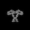
| Title | Cross-linked Contactin 2 Ig1-Ig6 | |||||||||
 Map data Map data | ||||||||||
 Sample Sample |
| |||||||||
 Keywords Keywords | contactins / adhesion molecule / protein structure / conformational changes / homodimer / MEMBRANE PROTEIN | |||||||||
| Function / homology |  Function and homology information Function and homology informationestablishment of protein localization to juxtaparanode region of axon / presynaptic membrane organization / regulation of axon diameter / positive regulation of adenosine receptor signaling pathway / L1CAM interactions / regulation of astrocyte differentiation / reduction of food intake in response to dietary excess / clustering of voltage-gated potassium channels / cerebral cortex GABAergic interneuron migration / dendrite self-avoidance ...establishment of protein localization to juxtaparanode region of axon / presynaptic membrane organization / regulation of axon diameter / positive regulation of adenosine receptor signaling pathway / L1CAM interactions / regulation of astrocyte differentiation / reduction of food intake in response to dietary excess / clustering of voltage-gated potassium channels / cerebral cortex GABAergic interneuron migration / dendrite self-avoidance / protein localization to juxtaparanode region of axon / NrCAM interactions / central nervous system myelination / cell-cell adhesion mediator activity / positive regulation of protein processing / G protein-coupled adenosine receptor signaling pathway / axon initial segment / NCAM1 interactions / juxtaparanode region of axon / node of Ranvier / regulation of cell morphogenesis / axonal fasciculation / adult walking behavior / negative regulation of neuron differentiation / fat cell differentiation / homophilic cell adhesion via plasma membrane adhesion molecules / regulation of neuronal synaptic plasticity / side of membrane / axon guidance / learning / establishment of localization in cell / synapse organization / protein processing / receptor internalization / microtubule cytoskeleton organization / myelin sheath / carbohydrate binding / postsynaptic membrane / cell adhesion / axon / neuronal cell body / synapse / cell surface / plasma membrane Similarity search - Function | |||||||||
| Biological species |  Homo sapiens (human) Homo sapiens (human) | |||||||||
| Method | single particle reconstruction / cryo EM / Resolution: 3.51 Å | |||||||||
 Authors Authors | Liu JL / Fan SF / Ren GR / Rudenko GR | |||||||||
| Funding support |  United States, 1 items United States, 1 items
| |||||||||
 Citation Citation | Journal: Structure / Year: 2024 Title: Molecular mechanism of contactin 2 homophilic interaction. Authors: Shanghua Fan / Jianfang Liu / Nicolas Chofflet / Aaron O Bailey / William K Russell / Ziqi Zhang / Hideto Takahashi / Gang Ren / Gabby Rudenko /  Abstract: Contactin 2 (CNTN2) is a cell adhesion molecule involved in axon guidance, neuronal migration, and fasciculation. The ectodomains of CNTN1-CNTN6 are composed of six Ig domains (Ig1-Ig6) and four FN ...Contactin 2 (CNTN2) is a cell adhesion molecule involved in axon guidance, neuronal migration, and fasciculation. The ectodomains of CNTN1-CNTN6 are composed of six Ig domains (Ig1-Ig6) and four FN domains. Here, we show that CNTN2 forms transient homophilic interactions (K ∼200 nM). Cryo-EM structures of full-length CNTN2 and CNTN2_Ig1-Ig6 reveal a T-shaped homodimer formed by intertwined, parallel monomers. Unexpectedly, the horseshoe-shaped Ig1-Ig4 headpieces extend their Ig2-Ig3 tips outwards on either side of the homodimer, while Ig4, Ig5, Ig6, and the FN domains form a central stalk. Cross-linking mass spectrometry and cell-based binding assays confirm the 3D assembly of the CNTN2 homodimer. The interface mediating homodimer formation differs between CNTNs, as do the homophilic versus heterophilic interaction mechanisms. The CNTN family thus encodes a versatile molecular platform that supports a very diverse portfolio of protein interactions and that can be leveraged to strategically guide neural circuit development. | |||||||||
| History |
|
- Structure visualization
- Downloads & links
-EMDB archive
| Map data | emd_44396.map.gz | 88.6 MB | EMDB map data format | |
|---|---|---|---|---|
| Header (meta data) | emd-44396-v30.xmlemd-44396.xml | 16.4 KB 16.4 KB | Display Display | EMDB header |
| FSC (resolution estimation) | emd_44396_fsc.xml | 10.6 KB | Display | FSC data file |
| Images |  emd_44396.png emd_44396.png | 38.6 KB | ||
| Filedesc metadata | emd-44396.cif.gz | 5.9 KB | ||
| Others | emd_44396_half_map_1.map.gzemd_44396_half_map_2.map.gz | 115.8 MB 115.8 MB | ||
| Archive directory |  http://ftp.pdbj.org/pub/emdb/structures/EMD-44396ftp://ftp.pdbj.org/pub/emdb/structures/EMD-44396 http://ftp.pdbj.org/pub/emdb/structures/EMD-44396ftp://ftp.pdbj.org/pub/emdb/structures/EMD-44396 | HTTPS FTP |
-Validation report
| Summary document | emd_44396_validation.pdf.gz | 840.1 KB | Display | EMDB validaton report |
|---|---|---|---|---|
| Full document | emd_44396_full_validation.pdf.gz | 839.7 KB | Display | |
| Data in XML | emd_44396_validation.xml.gz | 18.3 KB | Display | |
| Data in CIF | emd_44396_validation.cif.gz | 23.5 KB | Display | |
| Arichive directory | https://ftp.pdbj.org/pub/emdb/validation_reports/EMD-44396ftp://ftp.pdbj.org/pub/emdb/validation_reports/EMD-44396 | HTTPS FTP |
-Related structure data
| Related structure data | 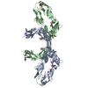 9ba5MC 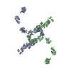 9ba4C M: atomic model generated by this map C: citing same article ( |
|---|---|
| Similar structure data |

-Links
| EMDB pages | EMDB (EBI/PDBe) / EMDataResource |
|---|---|
| Related items in Molecule of the Month |


-Map
| File | Download / File: emd_44396.map.gz / Format: CCP4 / Size: 125 MB / Type: IMAGE STORED AS FLOATING POINT NUMBER (4 BYTES) | ||||||||||||||||||||
|---|---|---|---|---|---|---|---|---|---|---|---|---|---|---|---|---|---|---|---|---|---|
| Voxel size | X=Y=Z: 1.05 Å | ||||||||||||||||||||
| Density |
| ||||||||||||||||||||
| Symmetry | Space group: 1 | ||||||||||||||||||||
| Details | EMDB XML:
|
-Supplemental data
- Sample components
-Entire : Contactin 2 Ig1-Ig6
| Entire | Name: Contactin 2 Ig1-Ig6 |
|---|---|
| Components |
|
-Supramolecule #1: Contactin 2 Ig1-Ig6
| Supramolecule | Name: Contactin 2 Ig1-Ig6 / type: organelle_or_cellular_component / ID: 1 / Parent: 0 / Macromolecule list: #1 |
|---|---|
| Source (natural) | Organism: Homo sapiens (human) |
-Macromolecule #1: Contactin-2
| Macromolecule | Name: Contactin-2 / type: protein_or_peptide / ID: 1 / Number of copies: 2 / Enantiomer: LEVO |
|---|---|
| Source (natural) | Organism: Homo sapiens (human) |
| Molecular weight | Theoretical: 65.394629 KDa |
| Recombinant expression | Organism:  Trichoplusia ni (cabbage looper) Trichoplusia ni (cabbage looper) |
| Sequence | String: ELGTGLESQT TFGPVFEDQP LSVLFPEEST EEQVLLACRA RASPPATYRW KMNGTEMKLE PGSRHQLVGG NLVIMNPTKA QDAGVYQCL ASNPVGTVVS REAILRFGFL QEFSKEERDP VKAHEGWGVM LPCNPPAHYP GLSYRWLLNE FPNFIPTDGR H FVSQTTGN ...String: ELGTGLESQT TFGPVFEDQP LSVLFPEEST EEQVLLACRA RASPPATYRW KMNGTEMKLE PGSRHQLVGG NLVIMNPTKA QDAGVYQCL ASNPVGTVVS REAILRFGFL QEFSKEERDP VKAHEGWGVM LPCNPPAHYP GLSYRWLLNE FPNFIPTDGR H FVSQTTGN LYIARTNASD LGNYSCLATS HMDFSTKSVF SKFAQLNLAA EDTRLFAPSI KARFPAETYA LVGQQVTLEC FA FGNPVPR IKWRKVDGSL SPQWTTAEPT LQIPSVSFED EGTYECEAEN SKGRDTVQGR IIVQAQPEWL KVISDTEADI GSN LRWGCA AAGKPRPTVR WLRNGEPLAS QNRVEVLAGD LRFSKLSLED SGMYQCVAEN KHGTIYASAE LAVQALAPDF RLNP VRRLI PAARGGEILI PCQPRAAPKA VVLWSKGTEI LVNSSRVTVT PDGTLIIRNI SRSDEGKYTC FAENFMGKAN STGIL SVRD ATKITLAPSS ADINLGDNLT LQCHASHDPT MDLTFTWTLD DFPIDFDKPG GHYRRTNVKE TIGDLTILNA QLRHGG KYT CMAQTVVDSA SKEATVLVRG SASTSHHHHH H UniProtKB: Contactin-2 |
-Macromolecule #2: 2-acetamido-2-deoxy-beta-D-glucopyranose
| Macromolecule | Name: 2-acetamido-2-deoxy-beta-D-glucopyranose / type: ligand / ID: 2 / Number of copies: 10 / Formula: NAG |
|---|---|
| Molecular weight | Theoretical: 221.208 Da |
| Chemical component information |  ChemComp-NAG: |
-Experimental details
-Structure determination
| Method | cryo EM |
|---|---|
 Processing Processing | single particle reconstruction |
| Aggregation state | particle |
-Sample preparation
| Concentration | 0.025 mg/mL | |||||||||
|---|---|---|---|---|---|---|---|---|---|---|
| Buffer | pH: 8 Component:
Details: 20 mM HEPES pH 8.0, 50 mM NaCl | |||||||||
| Vitrification | Cryogen name: ETHANE / Chamber humidity: 90 % / Chamber temperature: 281 K / Instrument: LEICA EM GP |
- Electron microscopy
| Microscope | TFS KRIOS |
|---|---|
| Specialist optics | Energy filter - Name: GIF Bioquantum / Energy filter - Slit width: 20 eV |
| Image recording | Film or detector model: GATAN K3 (6k x 4k) / Number grids imaged: 1 / Number real images: 4297 / Average exposure time: 7.39 sec. / Average electron dose: 50.0 e/Å2 |
| Electron beam | Acceleration voltage: 300 kV / Electron source:  FIELD EMISSION GUN FIELD EMISSION GUN |
| Electron optics | Illumination mode: FLOOD BEAM / Imaging mode: BRIGHT FIELD / Nominal defocus max: 1.6 µm / Nominal defocus min: 0.6 µm / Nominal magnification: 81000 |
| Sample stage | Specimen holder model: FEI TITAN KRIOS AUTOGRID HOLDER |
| Experimental equipment |  Model: Titan Krios / Image courtesy: FEI Company |
-Image processing
| Startup model | Type of model: NONE Details: The initial volume was generated by ab initio reconstruction. |
|---|---|
| Final reconstruction | Resolution.type: BY AUTHOR / Resolution: 3.51 Å / Resolution method: FSC 0.143 CUT-OFF / Software - Name: cryoSPARC (ver. 4.3.0) / Number images used: 98082 |
| Initial angle assignment | Type: RANDOM ASSIGNMENT / Software - Name: cryoSPARC (ver. 4.3.0) |
| Final angle assignment | Type: MAXIMUM LIKELIHOOD / Software - Name: cryoSPARC (ver. 4.3.0) |
-Atomic model buiding 1
| Refinement | Protocol: FLEXIBLE FIT |
|---|---|
| Output model | PDB-9ba5: |
