 Movie
Movie Controller


+ Open data

- Basic information
| Entry | 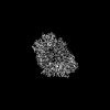 | |||||||||
|---|---|---|---|---|---|---|---|---|---|---|
| Title | Mycobacterium tuberculosis CoaX Homotetramer | |||||||||
 Map data Map data | ||||||||||
 Sample Sample |
| |||||||||
 Keywords Keywords | Pantothenate Kinase Isoform / CYTOSOLIC PROTEIN | |||||||||
| Function / homology |  Function and homology information Function and homology informationpantothenate kinase / pantothenate kinase activity / coenzyme A biosynthetic process / ATP binding / metal ion binding / cytoplasm Similarity search - Function | |||||||||
| Biological species |   Mycobacterium tuberculosis (bacteria) Mycobacterium tuberculosis (bacteria) | |||||||||
| Method | single particle reconstruction / cryo EM / Resolution: 2.71 Å | |||||||||
 Authors Authors | Chen J / Ekiert DC / Bhabha G | |||||||||
| Funding support |  United States, 2 items United States, 2 items
| |||||||||
 Citation Citation | Journal: Proc Natl Acad Sci U S A / Year: 2024 Title: Identification of a depupylation regulator for an essential enzyme in . Authors: Shoshanna C Kahne / Jin Hee Yoo / James Chen / Kehilwe Nakedi / Lakshminarayan M Iyer / Gregory Putzel / Nora M Samhadaneh / Alejandro Pironti / L Aravind / Damian C Ekiert / Gira Bhabha / ...Authors: Shoshanna C Kahne / Jin Hee Yoo / James Chen / Kehilwe Nakedi / Lakshminarayan M Iyer / Gregory Putzel / Nora M Samhadaneh / Alejandro Pironti / L Aravind / Damian C Ekiert / Gira Bhabha / Kyu Y Rhee / K Heran Darwin / Abstract: In , proteins that are posttranslationally modified with a prokaryotic ubiquitin-like protein (Pup) can be degraded by bacterial proteasomes. A single Pup-ligase and depupylase shape the pupylome, ...In , proteins that are posttranslationally modified with a prokaryotic ubiquitin-like protein (Pup) can be degraded by bacterial proteasomes. A single Pup-ligase and depupylase shape the pupylome, but the mechanisms regulating their substrate specificity are incompletely understood. Here, we identified a depupylation regulator, a protein called CoaX, through its copurification with the depupylase Dop. CoaX is a pseudopantothenate kinase that showed evidence of binding to pantothenate, an essential nutrient synthesizes, but not its phosphorylation. In a ∆ mutant, pantothenate synthesis enzymes including PanB, a substrate of the Pup-proteasome system (PPS), were more abundant than in the parental strain. In vitro, CoaX specifically accelerated depupylation of Pup~PanB, while addition of pantothenate inhibited this reaction. In culture, media supplementation with pantothenate decreased PanB levels, which required CoaX. Collectively, we propose CoaX regulates PanB abundance in response to pantothenate levels by modulating its vulnerability to proteolysis by proteasomes. | |||||||||
| History |
|
- Structure visualization
| Supplemental images |
|---|

- Downloads & links
-EMDB archive
| Map data | emd_44304.map.gz | 59.8 MB | EMDB map data format | |
|---|---|---|---|---|
| Header (meta data) | emd-44304-v30.xmlemd-44304.xml | 18.9 KB 18.9 KB | Display Display | EMDB header |
| FSC (resolution estimation) | emd_44304_fsc.xml | 8.4 KB | Display | FSC data file |
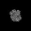
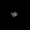
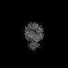
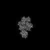
| Images |  emd_44304.png emd_44304.png | 87.8 KB | ||
| Filedesc metadata | emd-44304.cif.gz | 6.3 KB | ||
| Others | emd_44304_half_map_1.map.gzemd_44304_half_map_2.map.gz | 59.5 MB 59.5 MB | ||
| Archive directory |  http://ftp.pdbj.org/pub/emdb/structures/EMD-44304ftp://ftp.pdbj.org/pub/emdb/structures/EMD-44304 http://ftp.pdbj.org/pub/emdb/structures/EMD-44304ftp://ftp.pdbj.org/pub/emdb/structures/EMD-44304 | HTTPS FTP |
-Related structure data
| Related structure data | 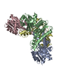 9b79MC 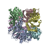 9b78C 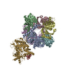 9ckuC C: citing same article ( M: atomic model generated by this map |
|---|---|
| Similar structure data |
-Links
| EMDB pages | EMDB (EBI/PDBe) / EMDataResource |
|---|
-Map
| File | Download / File: emd_44304.map.gz / Format: CCP4 / Size: 64 MB / Type: IMAGE STORED AS FLOATING POINT NUMBER (4 BYTES) | ||||||||||||||||||||||||||||||||||||
|---|---|---|---|---|---|---|---|---|---|---|---|---|---|---|---|---|---|---|---|---|---|---|---|---|---|---|---|---|---|---|---|---|---|---|---|---|---|
| Projections & slices | Image control
Images are generated by Spider. | ||||||||||||||||||||||||||||||||||||
| Voxel size | X=Y=Z: 0.826 Å | ||||||||||||||||||||||||||||||||||||
| Density |
| ||||||||||||||||||||||||||||||||||||
| Symmetry | Space group: 1 | ||||||||||||||||||||||||||||||||||||
| Details | EMDB XML:
|
 Z (Sec.)
 Y (Row.)
 X (Col.)


-Supplemental data
-Half map: #1
| File | emd_44304_half_map_1.map | ||||||||||||
|---|---|---|---|---|---|---|---|---|---|---|---|---|---|
| Projections & Slices |
| ||||||||||||
| Density Histograms |


-Half map: #2
| File | emd_44304_half_map_2.map | ||||||||||||
|---|---|---|---|---|---|---|---|---|---|---|---|---|---|
| Projections & Slices |
| ||||||||||||
| Density Histograms |


- Sample components
-Entire : Homotetrameric assembly of M. tuberculosis CoaX
| Entire | Name: Homotetrameric assembly of M. tuberculosis CoaX |
|---|---|
| Components |
|
-Supramolecule #1: Homotetrameric assembly of M. tuberculosis CoaX
| Supramolecule | Name: Homotetrameric assembly of M. tuberculosis CoaX / type: complex / ID: 1 / Parent: 0 / Macromolecule list: all |
|---|---|
| Source (natural) | Organism: Mycobacterium tuberculosis (bacteria) |
-Macromolecule #1: Type III pantothenate kinase
| Macromolecule | Name: Type III pantothenate kinase / type: protein_or_peptide / ID: 1 / Number of copies: 4 / Enantiomer: LEVO / EC number: pantothenate kinase |
|---|---|
| Source (natural) | Organism: Mycobacterium tuberculosis (bacteria) |
| Molecular weight | Theoretical: 29.341678 KDa |
| Recombinant expression | Organism: |
| Sequence | String: MLLAIDVRNT HTVVGLLSGM KEHAKVVQQW RIRTESEVTA DELALTIDGL IGEDSERLTG TAALSTVPSV LHEVRIMLDQ YWPSVPHVL IEPGVRTGIP LLVDNPKEVG ADRIVNCLAA YDRFRKAAIV VDFGSSICVD VVSAKGEFLG GAIAPGVQVS S DAAAARSA ...String: MLLAIDVRNT HTVVGLLSGM KEHAKVVQQW RIRTESEVTA DELALTIDGL IGEDSERLTG TAALSTVPSV LHEVRIMLDQ YWPSVPHVL IEPGVRTGIP LLVDNPKEVG ADRIVNCLAA YDRFRKAAIV VDFGSSICVD VVSAKGEFLG GAIAPGVQVS S DAAAARSA ALRRVELARP RSVVGKNTVE CMQAGAVFGF AGLVDGLVGR IREDVSGFSV DHDVAIVATG HTAPLLLPEL HT VDHYDQH LTLQGLRLVF ERNLEVQRGR LKTAR UniProtKB: Type III pantothenate kinase |
-Experimental details
-Structure determination
| Method | cryo EM |
|---|---|
 Processing Processing | single particle reconstruction |
| Aggregation state | particle |
-Sample preparation
| Concentration | 0.1 mg/mL | ||||||||||||
|---|---|---|---|---|---|---|---|---|---|---|---|---|---|
| Buffer | pH: 8 Component:
| ||||||||||||
| Grid | Model: Quantifoil R2/2 / Material: COPPER / Mesh: 300 / Support film - Material: CARBON / Support film - topology: CONTINUOUS / Support film - Film thickness: 2 | ||||||||||||
| Vitrification | Cryogen name: ETHANE / Chamber humidity: 100 % / Chamber temperature: 298 K / Instrument: FEI VITROBOT MARK IV |
- Electron microscopy
| Microscope | FEI TITAN KRIOS |
|---|---|
| Specialist optics | Energy filter - Slit width: 20 eV |
| Image recording | Film or detector model: GATAN K3 (6k x 4k) / Number grids imaged: 1 / Number real images: 7913 / Average exposure time: 1.8 sec. / Average electron dose: 48.51 e/Å2 |
| Electron beam | Acceleration voltage: 300 kV / Electron source:  FIELD EMISSION GUN FIELD EMISSION GUN |
| Electron optics | Illumination mode: FLOOD BEAM / Imaging mode: BRIGHT FIELD / Cs: 2.7 mm / Nominal defocus max: 2.5 µm / Nominal defocus min: 0.8 µm / Nominal magnification: 105000 |
| Sample stage | Specimen holder model: FEI TITAN KRIOS AUTOGRID HOLDER / Cooling holder cryogen: NITROGEN |
| Experimental equipment |  Model: Titan Krios / Image courtesy: FEI Company |


