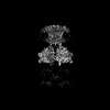
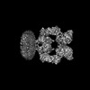
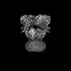
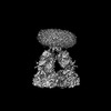
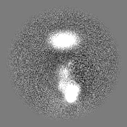
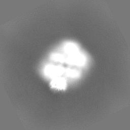
- EMDB-44125: Structure of concanavalin A (ConA) dimer from the open-state stru... -
+
Open data
ID or keywords:
Loading...
-
Basic information
Entry
Database: EMDB / ID: EMD-44125
Title
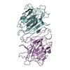
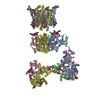
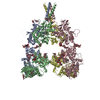
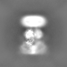
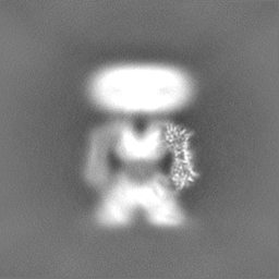
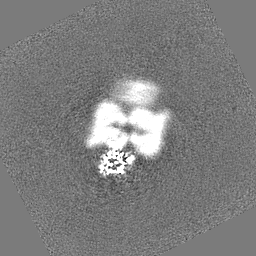
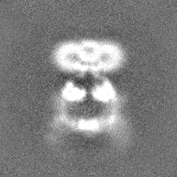
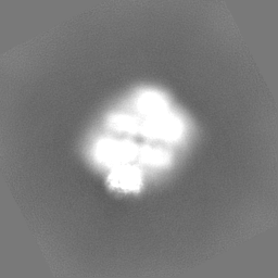
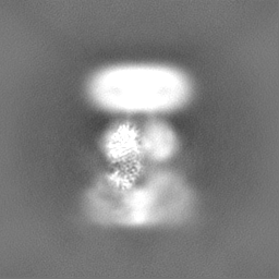
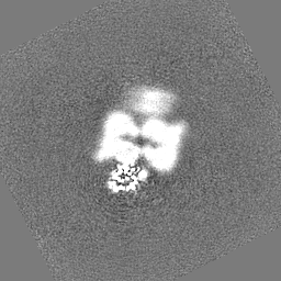
Structure of concanavalin A (ConA) dimer from the open-state structure of kainate receptor GluK2 in complex with agonist glutamate and positive allosteric modulator BPAM344 bound to two ConA dimers. Type I interface between GluK2 ligand-binding domain and ConA
Map data
Sample
Complex: Concanavalin-A dimer
Protein or peptide: Concanavalin A
Ligand: ZINC ION
Ligand: CALCIUM ION
Keywords
kainate receptor / GluK2 / glutamate / positive allosteric modulator / BPAM344 / open / concanavalin A / ConA / MEMBRANE PROTEIN
Function / homology
Function and homology information
regulation of defense response to virus / D-mannose binding / defense response / metal ion binding Similarity search - Function
National Institutes of Health/National Institute of Neurological Disorders and Stroke (NIH/NINDS)
R01 NS083660
United States
National Institutes of Health/National Institute of Neurological Disorders and Stroke (NIH/NINDS)
R01 NS107253
United States
National Institutes of Health/National Institute of Arthritis and Musculoskeletal and Skin Diseases (NIH/NIAMS)
R01 AR078814
United States
National Institutes of Health/National Cancer Institute (NIH/NCI)
R01 CA206573
United States
Citation
Journal: Nature / Year: 2024 Title: Kainate receptor channel opening and gating mechanism. Authors: Shanti Pal Gangwar / Maria V Yelshanskaya / Kirill D Nadezhdin / Laura Y Yen / Thomas P Newton / Muhammed Aktolun / Maria G Kurnikova / Alexander I Sobolevsky / Abstract: Kainate receptors, a subclass of ionotropic glutamate receptors, are tetrameric ligand-gated ion channels that mediate excitatory neurotransmission. Kainate receptors modulate neuronal circuits and ...Kainate receptors, a subclass of ionotropic glutamate receptors, are tetrameric ligand-gated ion channels that mediate excitatory neurotransmission. Kainate receptors modulate neuronal circuits and synaptic plasticity during the development and function of the central nervous system and are implicated in various neurological and psychiatric diseases, including epilepsy, depression, schizophrenia, anxiety and autism. Although structures of kainate receptor domains and subunit assemblies are available, the mechanism of kainate receptor gating remains poorly understood. Here we present cryo-electron microscopy structures of the kainate receptor GluK2 in the presence of the agonist glutamate and the positive allosteric modulators lectin concanavalin A and BPAM344. Concanavalin A and BPAM344 inhibit kainate receptor desensitization and prolong activation by acting as a spacer between the amino-terminal and ligand-binding domains and a stabilizer of the ligand-binding domain dimer interface, respectively. Channel opening involves the kinking of all four pore-forming M3 helices. Our structures reveal the molecular basis of kainate receptor gating, which could guide the development of drugs for treatment of neurological disorders.
Name: ZINC ION / type: ligand / ID: 2 / Number of copies: 2 / Formula: ZN
Molecular weight
Theoretical: 65.409 Da
-
Macromolecule #3: CALCIUM ION
Macromolecule
Name: CALCIUM ION / type: ligand / ID: 3 / Number of copies: 2 / Formula: CA
Molecular weight
Theoretical: 40.078 Da
-
Experimental details
-
Structure determination
Method
cryo EM
Processing
single particle reconstruction
Aggregation state
particle
-
Sample preparation
Buffer
pH: 8 Component:
Concentration
Formula
Name
150.0 mM
NaCl
sodium chloride
20.0 mM
tris(hydroxymethyl)aminomethane
1.0 mM
beta-Mercaptoethanol
0.05 %
digitonin
0.5 mM
BPAM344
0.09 mM
concanavalin A
2.5 mM
L-Glutamic acid
Grid
Model: UltrAuFoil R1.2/1.3 / Material: GOLD / Support film - Material: GOLD / Support film - topology: HOLEY
Vitrification
Cryogen name: ETHANE
-
Electron microscopy
Microscope
TFS KRIOS
Image recording
Film or detector model: GATAN K3 (6k x 4k) / Digitization - Dimensions - Width: 5760 pixel / Digitization - Dimensions - Height: 4092 pixel / Number grids imaged: 5 / Number real images: 22990 / Average exposure time: 2.5 sec. / Average electron dose: 58.0 e/Å2
Electron beam
Acceleration voltage: 300 kV / Electron source: FIELD EMISSION GUN
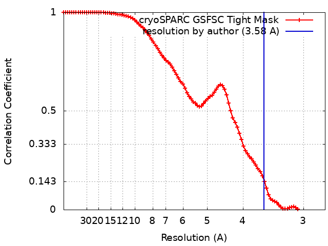
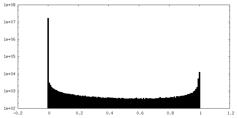
Resolution.type: BY AUTHOR / Resolution: 3.58 Å / Resolution method: FSC 0.143 CUT-OFF / Software - Name: cryoSPARC (ver. 4.4.1) / Number images used: 163519
Initial angle assignment
Type: OTHER / Software - Name: cryoSPARC (ver. 4.4.1)
Final angle assignment
Type: OTHER / Software - Name: cryoSPARC (ver. 4.4.1)
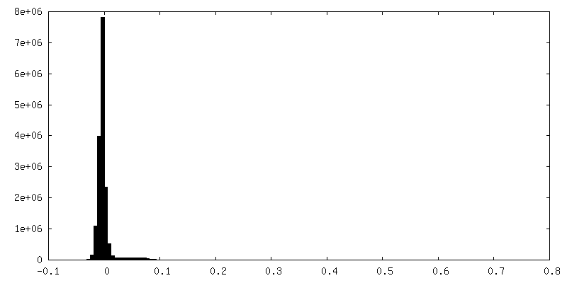
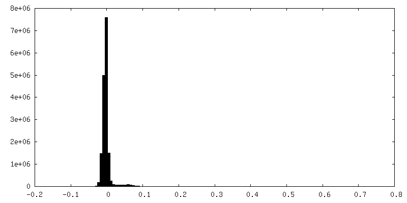
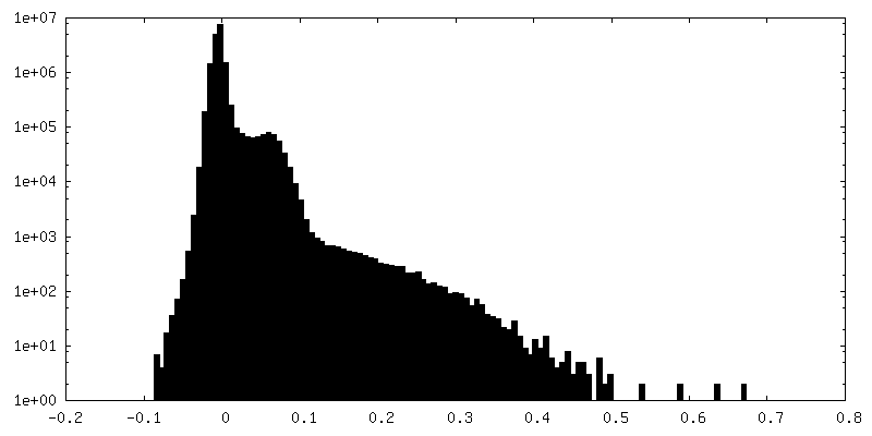
FSC plot (resolution estimation)
-
Atomic model buiding 1
Refinement
Space: REAL
Output model
PDB-9b34: Structure of concanavalin A (ConA) dimer from the open-state structure of kainate receptor GluK2 in complex with agonist glutamate and positive allosteric modulator BPAM344 bound to two ConA dimers. Type I interface between GluK2 ligand-binding domain and ConA
+
About Yorodumi
-
News
-
Feb 9, 2022. New format data for meta-information of EMDB entries
New format data for meta-information of EMDB entries
Version 3 of the EMDB header file is now the official format.
The previous official version 1.9 will be removed from the archive.
In the structure databanks used in Yorodumi, some data are registered as the other names, "COVID-19 virus" and "2019-nCoV". Here are the details of the virus and the list of structure data.
Jan 31, 2019. EMDB accession codes are about to change! (news from PDBe EMDB page)
EMDB accession codes are about to change! (news from PDBe EMDB page)
The allocation of 4 digits for EMDB accession codes will soon come to an end. Whilst these codes will remain in use, new EMDB accession codes will include an additional digit and will expand incrementally as the available range of codes is exhausted. The current 4-digit format prefixed with “EMD-” (i.e. EMD-XXXX) will advance to a 5-digit format (i.e. EMD-XXXXX), and so on. It is currently estimated that the 4-digit codes will be depleted around Spring 2019, at which point the 5-digit format will come into force.
The EM Navigator/Yorodumi systems omit the EMD- prefix.
Related info.:Q: What is EMD? / ID/Accession-code notation in Yorodumi/EM Navigator
Yorodumi is a browser for structure data from EMDB, PDB, SASBDB, etc.
This page is also the successor to EM Navigator detail page, and also detail information page/front-end page for Omokage search.
The word "yorodu" (or yorozu) is an old Japanese word meaning "ten thousand". "mi" (miru) is to see.
Related info.:EMDB / PDB / SASBDB / Comparison of 3 databanks / Yorodumi Search / Aug 31, 2016. New EM Navigator & Yorodumi / Yorodumi Papers / Jmol/JSmol / Function and homology information / Changes in new EM Navigator and Yorodumi
 Movie
Movie Controller

 Yorodumi
 Open data
 Basic information
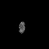
 Map data
 Sample
 Keywords
 Function and homology information
 Canavalia ensiformis (jack bean)
 Authors
 United States, 4 items
  Citation
 Structure visualization
 Downloads & links
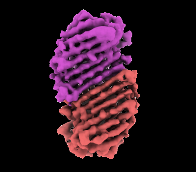 emd_44125.png
 http://ftp.pdbj.org/pub/emdb/structures/EMD-44125
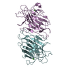


 Z (Sec.)
 Y (Row.)
 X (Col.)


 Sample components
 Processing
 Electron microscopy
 FIELD EMISSION GUN